1309
Readout-Segmented EPI with 2D-PACE for the Abdominal Diffusion Weighted Imaging1Siemens Shenzhen Magnetic Resonance Ltd, Shenzhen, People's Republic of China
Synopsis
The 2D-PACE technique has been proved to be superior to conventional respiratory triggering techniques for imaging the abdominal region because it could detect more accurate respiratory waveform, which is already widely used in single shot EPI for high quality abdominal diffusion weighted imaging. The readout-segmented EPI features much shorter echo spacing compared with single shot EPI and consequently enjoys less distortion. In this study, the 2D-PACE technique is integrated into a readout-segmented EPI sequence and makes high resolution abdominal diffusion images with less distortion and image blurring.
Purpose
Abdominal diffusion weighted imaging (DWI) is a valuable adjunct to traditional techniques and improves the sensitivity for both detection and characterization of disease processes. However it is important to prevent the respiratory motion as much as possible due to the strong effect of breathing on tissue displacement between expiration and inspiration, especially for the upper abdominal imaging. High signal averages or number of excitations are generally used for free-breathing single-shot EPI (ss-EPI) sequence to reduce the motion artifacts but will lead to image blurring. Moreover different techniques have been proposed to improve abdominal diffusion image quality by controlling or monitoring the respiratory movement, such as breath-hold, respiratory-belt (RESP-B)1 and 2D navigator-triggered prospective acquisition correction (2D-PACE)2. The breath-hold diffusion sequence is faster than triggered ones but will limit the spatial resolution due to the demand of high speed acquisitions and also require the patient cooperation. In addition, the registration among images acquired during different breath holds is necessary sometimes because the different respiratory depth in each breath hold. The 2D-PACE technique has been proved to be superior to conventional respiratory triggering techniques for imaging the abdominal region because it’s able to detect more accurate respiratory waveform3-4, which is already widely used in ss-EPI for high quality abdominal diffusion weighted (DW) images5.
Readout-segmented EPI (rs-EPI) allows a substantial reduction in echo spacing and enjoys a low level of distortion and blurring caused by T2* decay6 in comparison to ss-EPI, which has been validated in non-triggering applications, such as in head, spine and breast. In this study, we demonstrate how 2D-PACE technique can be used in a rs-EPI sequence and provide high resolution abdominal diffusion images with less distortion and image blurring.
Methods
The 2D-PACE technique is used to generate a non-product version of a commercial rs-EPI sequence (RESOLVE, Siemens Healthineers), referred as RESOLVE-PACE. The study was performed on a commercial 1.5T scanner (MAGNETOM Aera, Siemens Healthineers) equipped with a 20-channel headneck and a 32-channel spine coil. DW images from healthy volunteers were acquired with free breathing ss-EPI, RESOLVE, RESOLVE-RESP-B and RESOLVE-PACE protocols respectively. Imaging parameters were as follows: FOV = 306x380 mm2, matrix size = 108x134, 30 slices with 6mm thickness, in-plane GRAPPA factor = 2, b = 50/400/800 s/mm2, diffusion mode = 4-Scan Trace. The acquisition time for ss-EPI w/o and with 2D-PACE is 3:18 min and approx.4 min respectively, for RESOLVE w/o and with 2D-PACE is 3:29 min and approx.5 min, for RESOLVE-RESP-B is approx.4 min. Three readout segments with readout partial Fourier factor 6/8 was used for the protocols based on RESOLVE. An additional RESOLVE-PACE acquisition with matrix size 128x160 (approx. TA = 6min) was also performed as a higher resolution DWI. To save the scan time, the averages for different b values in RESOLVE are less than those in ss-EPI because RESOLVE usually has a higher SNR compared with ss-EPI.Results
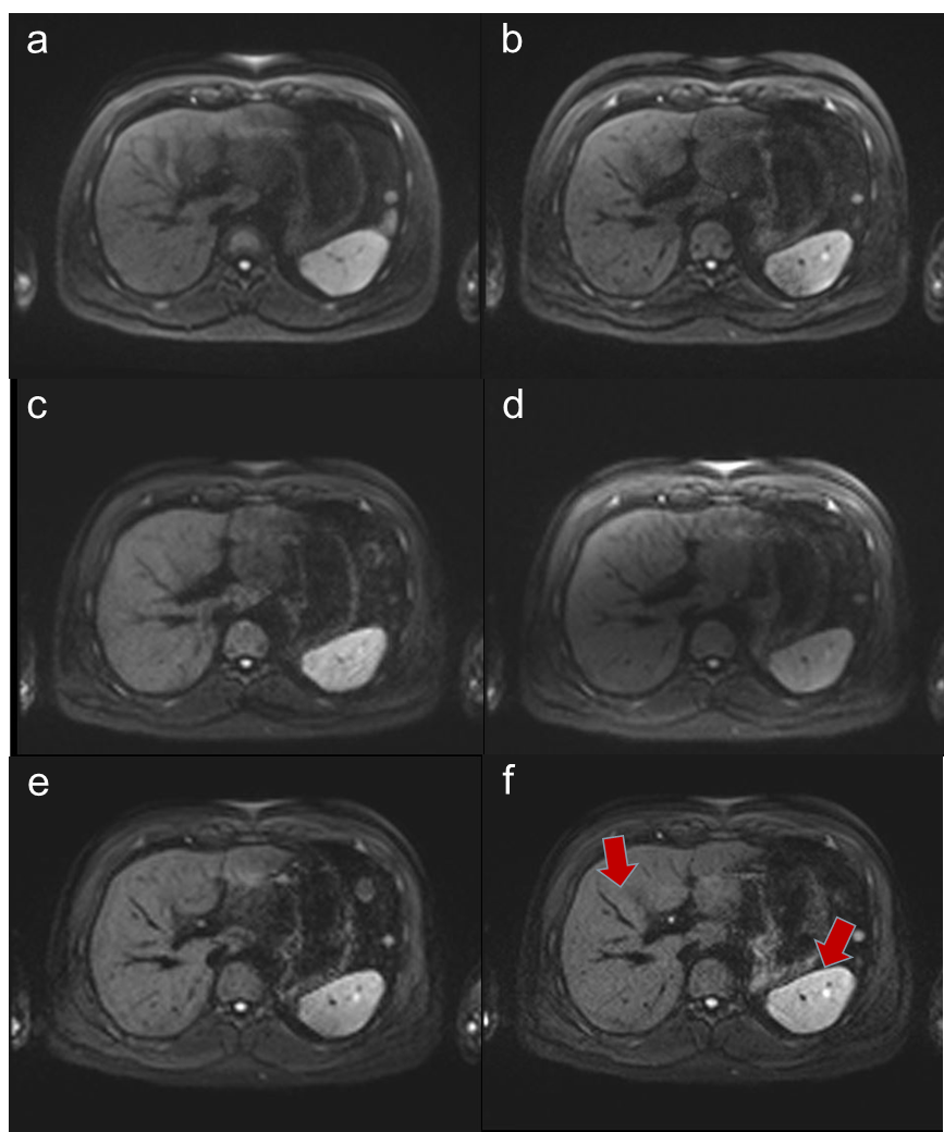
Fig. 1 compares the DW sample images (b = 800 s/mm2) acquired using ss-EPI w/o and with 2D-PACE, RESOLVE-RESP-B, RESOLVE w/o and 2D-PACE at the similar slice position. Fig.1a-b are the images from ss-EPI w/o and with 2D PACE. Fig.1c-e are the images from RESOLVE with free breathing, RESP-B and 2D-PACE respectively. RESOLVE-PACE with a higher resolution is shown in Fig.1f. The motion induced artifacts and image blurring are much improved with triggering (Fig.1b, d-f) compare with free-breathing (Fig.1a, c). Fig.1f shows more clear structural details because of a higher resolution (see arrows in Fig.1f), and also enjoys the best image quality, considering the motion artifacts, imaging blurring and distortions.Discussion and Conclusion
We have demonstrated that the RESOLVE integrated with 2D-PACE could reduce respiratory motion artifact for abdominal DW images and improve image quality, in comparison to ss-EPI and conventional RESOLVE. The 2D-PACE acquisitions using ss-EPI and RESOLVE with the same resolution show comparable structure details but the later one should have reduced distortion due to the shorter echo spacing, which could be more obvious in the abdominal images acquired on 3T scanner. It notes that RESOLVE-PACE with a higher resolution could provide an increase level of structural detail without the cost of distortion and T2* blurring, which is promising for high resolution abdominal DWI.Acknowledgements
We thank for Alto Stemmer for his assistance with the 2D-PACE implementation on RESOLVE sequence.References
1. EhMan RL et al. Magnetic resonance imaging with respiratory gating: techniques and advantage. Am J Roentqenol. 1984;143(6):1175-82
2. Asbach P et al. Magnetic resonance cholangiopancreatography using a free-breathing T2-weighted turbo spin-echo sequence with navigator triggered prospective acquisition correction. Magn Reson Imaging. 2005;23:939 –945
3. Bong Soo Kim et al. Comparison of Three Free-Breathing T2-Weighted MRI Sequences in the Evaluation of Focal Liver Lesions. Am J Roentqenol. 2008; 190:19-27
4. Klessen C et al. Magnetic resonance imaging of the upper abdomen using a free-breathing T2-weighted turbo spin echo sequence with navigator triggered prospective acquisition correction. J Magn Reson Imaging. 2005; 21:576 –582
5. Bachir Taouli et al, Diffusion-Weighted Imaging of the Liver: Comparison of Navigator Triggered and Breathhold Acquisitions. J Magn Reson Imaging. 2008; 30:561-568
6. Porter D et al, High Resolution Diffusion-Weighted Imaging Using Readout-Segmented Echo-Planar Imaging, Parallel Imaging and a Two-Dimensional Navigator-Based Reacquisition, Magn Reson Imaging. 2009; 62:468-475
Figures