1298
Measuring the accuracy of prospective motion correction through retroactive application of estimates1Neuroradiology, Karolinska University Hospital, Stockholm, Sweden, 2Department of Clinical Neuroscience, Karolinska Institutet, Stockholm, Sweden
Synopsis
Several prospective motion correction methods have been presented to address the issue of motion induced artifacts in MRI. We present a method to determine the accuracy of prospective motion correction methods by applying estimates to a PROPELLER reconstruction.
Purpose
Several prospective motion correction methods have been presented to address the issue of motion induced artifacts in MRI. Since prospective methods alter the acquisition depending on the motion estimates, it is however difficult to compare different prospective navigator variants as the same patient motion cannot be reliably reproduced. Recently, we have presented a projection based fat navigator (1). As the projection based FatNav data is EPI based and of low resolution, both voxel size and geometric distortions are potential sources of inaccuracy in motion estimates even though the EPI acquisition is accelerated. Also, it is important to avoid a mixture of fat and water content, why the choice of RF excitation pulse may also affect the estimates. Therefore, we have investigated how the type of RF pulse and GRAPPA acceleration affects the collapsed FatNav motion estimates. This was done by performing a stepwise motion experiment, acquiring multiple variants of the FatNav navigators for each head pose in conjunction with a PROPELLER (2) acquisition and store the navigator data for offline retrospective correction.Methods
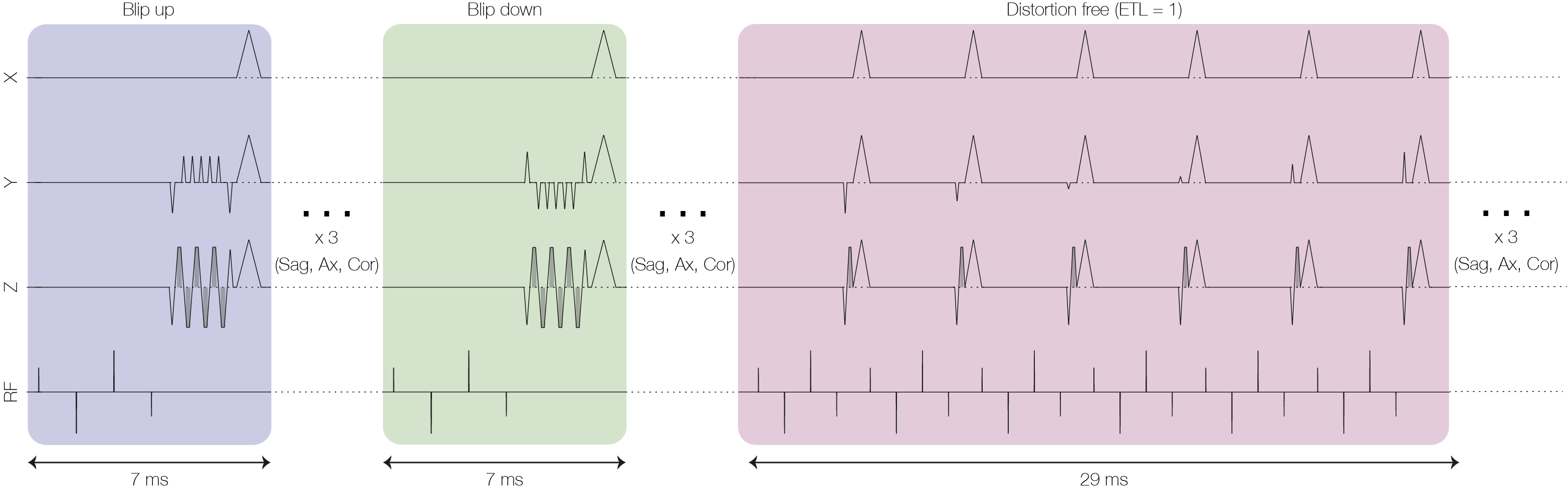
Controlled motion experiments were performed by trained volunteers, where the head was moved only between blade volumes. In these experiments, our 3D rigid body motion correction between blade volumes was expected to work optimally as there was no intra-blade motion. In the first experiment, the effect of geometric distortions on the accuracy of our projection-based FatNav motion estimates was investigated by playing out three projection FatNavs before the stack of slices for each blade volume for later offline use. The first two FatNavs were acquired with positive and negative blips, respectively, leading to geometric distortions in opposite directions. The third navigator was acquired with ETL=1 using several RF pulses to produce distortion free collapsed FatNav data for each blade volume. The sequence is illustrated in Figure 1. To allow for stepwise head motion between blade volumes, a two-second delay was inserted after each blade volume, before the FatNavs belonging to the next blade volume were played out. The motion correction part of the reconstruction was modified to optionally apply external motion estimates instead of those determined from the propeller data itself. Reconstructed images using the three sets (blipup, blipdown, distortion-free) of FatNav estimates were compared with estimated derived from the propeller blade data (3).
In a second experiment, three FatNavs were acquired before each propeller blade stack, but this time using the same EPI readouts (R=6, 36x36). The difference between the three navigators was instead the spectral RF pulse type; Minimum phase Fat Sat (MPFS), Binominal RF with 4 sub pulses (Bin4) and, Binomial RF with 8 sub pulses (Bin8).
The experiments were performed on a volunteer on a 3T clinical MRI system (DVMR750w, GE Healthcare, Milwaukee, WI) using a multi-channel receive coil (GEM Head and Neck Unit) and the following scan parameters for the PROPELLER acquisition: TE = 98 ms, 14 blades, matrix size = 320x32, 14 adjacent slices with 5 mm thickness, R=2 (cross-calibrated GRAPPA), and ETL=16. FatNav was acquired with a 30cm FOV, flip angle = 5 degrees, matrix size = 36x36. Bin8 RF pulse was used for the distortion experiment.
Results
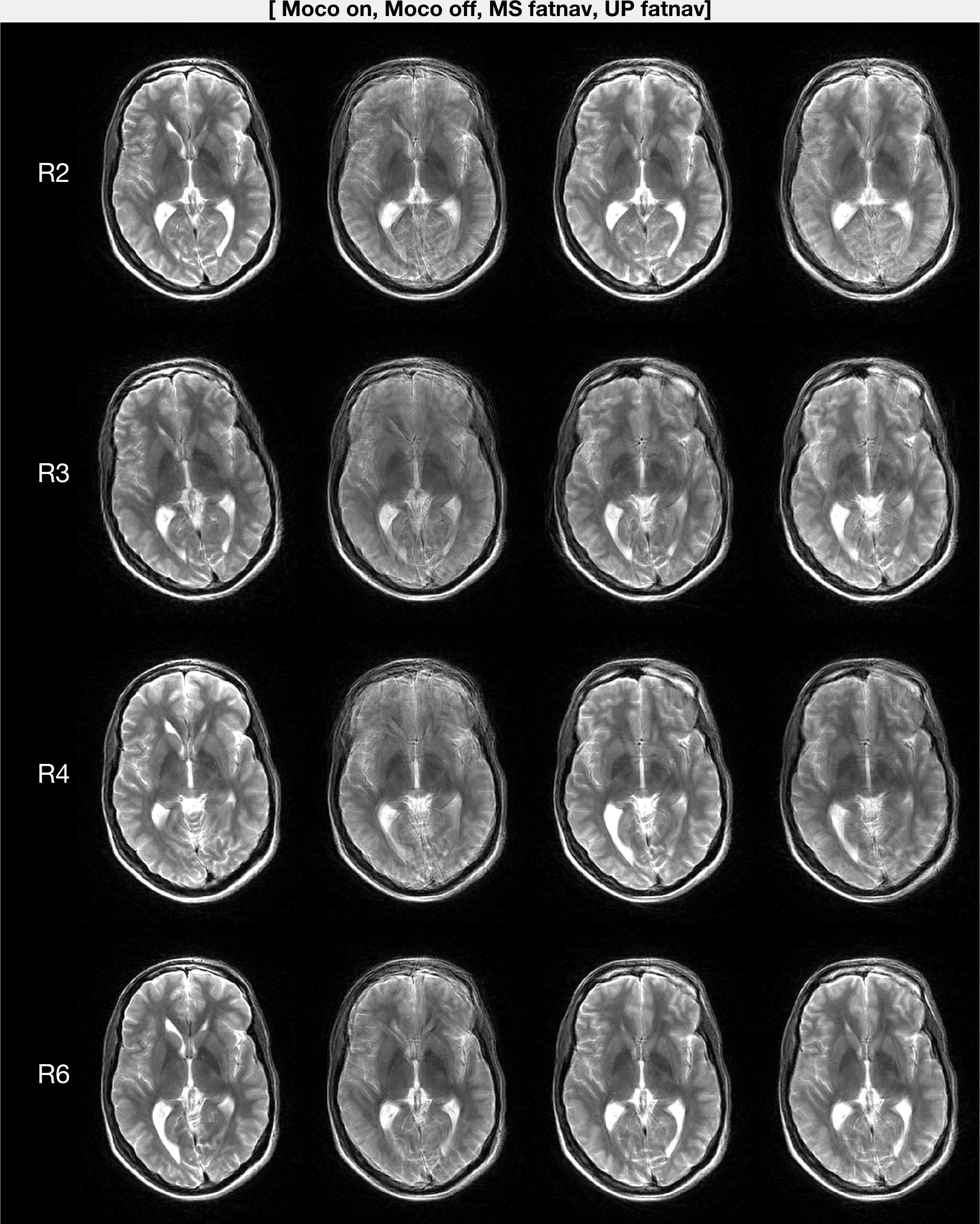
Figure 2 shows images reconstructed with FatNav estimates from four in-plane acceleration factors, as well the distortion-free FatNav. Increased in-plane acceleration yields more accurate estimates.
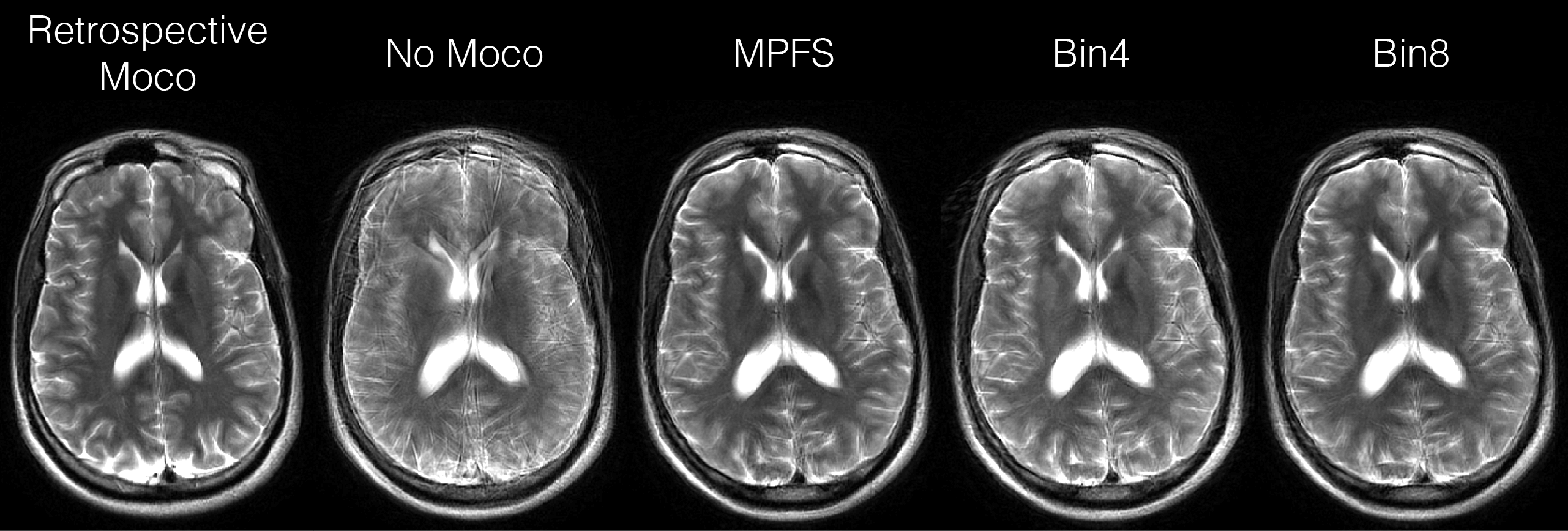
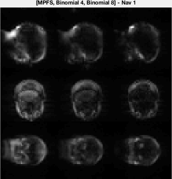
Figure 3 shows images reconstructed with distortion-free and R=6 FatNav using different RF pulses. A binomial fat-selective with 4 sub pulses (Bin4) the minimum-phase fat-selective (MPFS) pulse was compared using a flip angle of 5 degrees. The projection images from the distortion-free FatNav using Bin4, Bin8, and MPFS pulses are shown in Figure 4. Bin8 is preferable to both Bin4 and MPFS in terms of clean fat excitation of the scalp. Both MPFS and Bin4 excites the neck tissue which is not rigidly coupled to the head.
Discussion & Conclusion
While lower acceleration factors are less susceptible to parallel imaging artifacts, they are more distorted due to the decreased pseudo bandwidth. The distortion-free FatNav managed to track the head movement in all four scans. An increase in GRAPPA acceleration yields better estimates, and R=6 estimates were almost identical to the distortion-free estimates. The results also show that the choice of RF matters in terms of water leakage, but more work is needed to confirm potential differences in the accuracy of the motion estimates. In conclusion, we have presented a method that utilizes stepwise inter-blade motion with our PROPELLER sequence allowing different variants of navigators to be inserted before each change of head pose, and compared offline.Acknowledgements
No acknowledgement found.References
1. Avventi E, Engström M, Norbeck O, Mårtensson M, Skare S. Projection-based 2D/3D registration of collapsed FatNav data for prospective motion correction. In: Proc. Intl. Soc. Mag. Reson. Med. 23 (2015).
2. Pipe JG. Motion correction with PROPELLER MRI: application to head motion and free-breathing cardiac imaging. Magn. Reson. Med. 1999;42:963–969.
3. Skare S, Avventi E, Mårtensson M, Norbeck O, Engström M, Sandell M, Wang C. Propeller techniques for pediatric exams in the presence of large motion. In: Proc. Intl. Soc. Mag. Reson. Med. 23 (2015). Karolinska University Hospital; 2015.
Figures