1285
Golden-angle Sparse Liver Imaging: Radial or Cartesian Sampling?1Center for Advanced Imaging Innovation and Research (CAI2R), New York University School of Medicine, New York, NY, United States, 2Siemens Healthineers, New York, USA
Synopsis
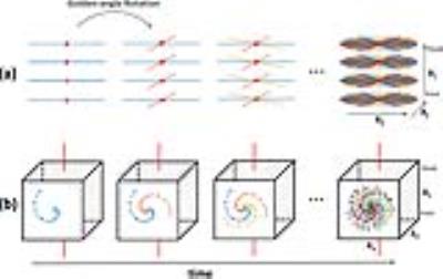
This work compares golden-angle stack-of-stars sampling and golden-angle Cartesian sampling for free-breathing liver MRI with eXtra-Dimensional (XD) compressed sensing reconstruction. For Cartesian sampling, the phase-encoding steps in the ky-kz plane are segmented into multiple interleaves that rotate at a golden angle. Each interleave starts from the center (ky=kz=0) of k-space and follows a pseudo-radial pattern on a Cartesian grid. Results from this initial study suggest that golden-angle Cartesian sampling achieves higher effective spatial resolution than radial sampling, but it still suffers from residual ghosting artifacts due to respiratory motion for free-breathing liver imaging.
INTRODUCTION
Golden-angle radial sampling1 has several attractive benefits for abdominopelvic imaging. It enables continuous data acquisitions and provides a high level of incoherence for the application of compressed sensing techniques2,3. The repeated sampling of the k-space center reduces sensitivity to motion4,5 and provides self-navigation6,7. However, compared to conventional Cartesian imaging, radial sampling requires longer reconstruction time and poses several challenges, including sensitivity to off-resonances, gradient delays, and non-ideal gradient-amplifier responses. These system imperfections may result in image distortion or blurring if not corrected properly. In recent years, several studies have incorporated the golden-angle rotation scheme into 3D Cartesian sampling8-12. Here, phase-encoding steps in ky-kz plane are segmented into multiple radial- or spiral-like interleaves, and each interleave is rotated by the golden angle from previous one. Such a sampling strategy is expected to overcome the challenges presented in radial imaging while offering the benefits of golden-angle radial sampling. Moreover, it provides higher flexibility in the design of the sampling trajectory, allowing either isotropic or anisotropic data acquisitions along different spatial dimensions. In this work, golden-angle radial and golden-angle Cartesian sampling schemes were compared for eXtra-Dimensional (XD) sparse reconstruction of liver MRI.METHODS
Sampling Trajectory Design: Our sampling trajectories were designed for axial acquisitions, which is the major imaging orientation employed in routine clinical liver MR exams. Golden-angle radial sampling was implemented using a star-of-stars trajectory, where radial encoding was employed in the kx-ky plane and fully-sampled Cartesian encoding was employed along the kz dimension (the head-to-foot direction in our study) (Figure1a). Respiratory motion signal was detected from the centers of k-space in each kx-ky plane (red dots), as described in (7). Golden-angle Cartesian sampling was implemented in the ky-kz plane, which are the anterior-posterior direction (ky) and the head-to-foot direction (kz), respectively (Figure1b). In addition, an extra k-space measurement orientated along kz (red lines in Figure1b) was consistently acquired in the end of each interleave for respiratory motion detection. The motion detection procedure was the same as that performed in radial imaging.
Imaging Studies: IRB-approved liver imaging was performed in 3 patients after contrast injection and also in one volunteer without intravenous contrast. For each subject, both golden-angle radial sampling and golden-angle Cartesian sampling were performed, and each dataset was continuously acquired for 90 seconds in a transverse orientation. Each dataset was sorted into four respiratory states using extracted motion signals, and motion-resolved sparse reconstruction (XD-Sparse) was performed as previously described in (7). For each pair of results (one radial and one Cartesian), a radiologist blinded to the acquisition schemes was asked to select a preferred result.
RESULTS
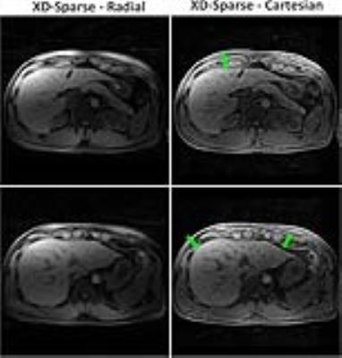
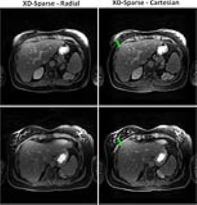
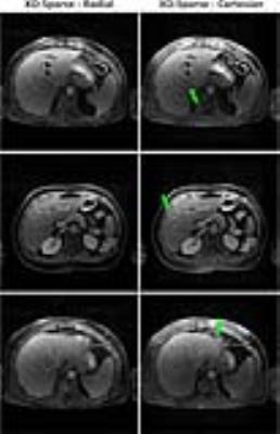
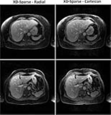
Figure2a shows non-contrast results from the volunteer comparing radial sampling (left) and Cartesian sampling (right) for two slices. These results suggest that radial sampling suffers from slight blurring, which is likely due to a combination of residual motion and off-resonance effects. Cartesian sampling, on the other hand, achieved increased image sharpness, but suffered from residual ghosting artifacts (green arrow) due to motion. For the first patient study in Figure3, radial sampling and Cartesian sampling achieved comparable image sharpness, but the latter suffered from residual ghosting artifacts (green arrows) as in the non-contrast case in the volunteer. The results from the second patient study (Figure4) were similar to those for the first patient, with residual ghosting artifacts clearly visible in the Cartesian approach. In the last patient study (Figure5), radial sampling and Cartesian sampling achieved comparable image sharpness, and motion artifacts were not noted in any of them, which is likely due to lighter breathing of the subject during data acquisition. For all the five pairs of images, a radiologist blinded to the acquisition scheme preferred the radial result.DISCUSSION
In this initial study, golden-angle Cartesian sampling produced slightly higher image sharpness, which is likely due to its immunity to system imperfections such as off-resonance and gradient-nonlinearity. However, the performance of radial sampling was more consistent and more reliable for free-breathing liver imaging due to its higher robustness to motion. Although the center of k-space is oversampled in both golden-angle radial and Cartesian trajectories, leading to reduced sensitivity to respiratory motion, the interpolation process in the gridding operation results in increased averaging effects in radial image reconstruction, which explains the appearance of residual ghosting artifacts in Cartesian sampling. However, although our study suggested that golden-angle radial sampling is more reliable for liver imaging, golden-angle Cartesian sampling could be a better choice for applications where motion is less of a challenge, such as DCE-MRI of the brain11 or pediatric imaging with sedation8,13Acknowledgements
This work was supported in part by the NIH, and was performed under the rubric of the Center for Advanced Imaging Innovation and Research (CAI2R), a NIBIB Biomedical Technology Resource Center (NIH P41 EB017183).References
[1] Winkelmann S et al. IEEE TMI 2007;26:68–76 [2] Block KT et al. MRM 2007;57:1086–1098 [4] Feng L et al. MRM 2014 72 (3), 707-717 [6] Glover GH et al. MRM 1992;28:275–289 [5] Chandarana H et al. Invest Radiol 2011;46:648–653 [6] Liu J et al. MRM 2010;63(5):1230-7 [7] Feng L et al. MRM 2016;75 (2), 775-788 [8] Cheng JY et al. JMRI 2015;Aug;42(2):407-20 [9] Liu J et al. Quantitative imaging in medicine and surgery 2014;4(1):57-67 [10] Prieto C et al. JMRI 2015;41(3):738-746 [11] Zhu Y et al. MRI 2015;34(7):940-950 [12] Forman C et al. Med Image Comput Comput Assist Interv. 2013;16(Pt 2):575-82 [13] Han F et al. MRM 2016 2016 Aug 16. doi: 10.1002/mrm.26376. [Epub ahead of print]Figures
