1275
Validating the accuracy and effectiveness of Prospective Motion Correction on rsfMRI1Cognition and Brain Science Unit, MRC, Cambridge, United Kingdom
Synopsis
Prospective motion correction for MRI has been shown to greatly improve image quality for structural scans but its impact on fMRI data is still unclear. In this work, we studied the effectiveness of prospective motion correction by analysing the accuracy of the tracking system and looking at the effects of motion correction on resting-state fMRI with no instructed subject motion.
Introduction
Advancements in magnetic field strength and imaging techniques have led to increasing resolution in function Magnetic Resonance Imaging (fMRI) studies. However, subject motion remains a significant confounding factor in attempts to reach even higher resolution. Numerous solutions have been proposed1, of which prospective motion correction (PMC) using optical trackers is one of the most promising. However, most of these studies have looked at structural data2, with very few focusing on fMRI. Moreover, past studies on fMRI data3 have used motion conditions where subjects were actively instructed to move. This is not necessarily representative of real motion patterns in actual studies where participants are instructed to remain as still as possible. Thus, the benefits of PMC reported by past studies could have been overestimated.
The PMC system employed in this study utilizes an in-bore optical camera to track a Moiré pattern marker, which provides positional information for all 6 degrees of freedom. Firstly, we attempt to validate the effectiveness of an optical PMC system by comparing the accuracy of the PMC system for different marker positions on the subject’s face. This will be followed by a resting state fMRI study of 18 subjects without active motion.
Methods
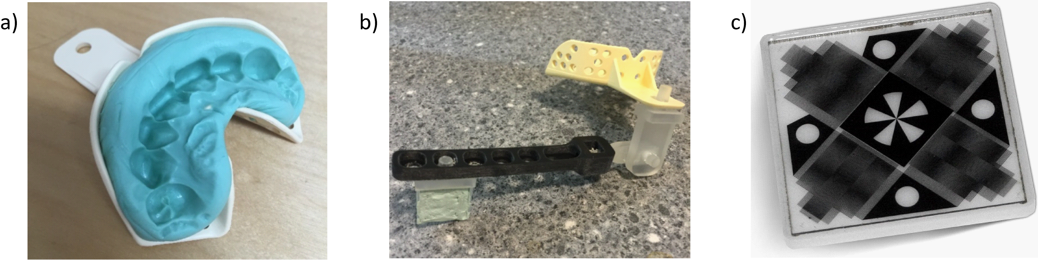
Preliminary tests on marker adhesion were carried out on 3 subjects. Subjects were instructed to remain as still as possible or to move their head at regular intervals for different scans. For comparison, the optical marker was attached to either the cheek or to a mouthpiece (Figure 1). The mouthpiece was custom-moulded on the spot for each subject by asking the subject to bite down on a dental putty for 2 minutes. Scan parameters of the preliminary EPI were: TR=1260ms TE=30ms FA=78o Matrix size=64*64*20 ToA=~5mins.
A subsequent resting state study was carried out on 18 participants. The subjects were required to be present for 3 separate scan sessions: (Condition 1) with motion correction and mouthpiece on, (Condition 2) with the mouthpiece on but motion correction switched off and (Condition 3) with no mouthpiece. Subject feedback on the mouthpiece was collected via a questionnaire after each session. The order of the three sessions was randomised across the 18 subjects. In all cases, subjects were told to remain as still as possible. This replicated the instructions usually given for fMRI studies, hence allowing us to assess the effect of PMC when only involuntary motion is present. Scan parameters of the resting state fMRI were: TR=2000ms TE=30ms FA=78o Matrix size=64*64*32 ToA=~8mins.
All scans were conducted on a 3T Prisma, Siemens, using a 32-channel head coil. Motion data were acquired using a commercially available optical PMC system (KinetiCor Inc, HI, USA). Most of the data analysis was done in SPM 8 and the realignment parameters were used to compare marker adhesion. Brain extraction was done using BET in FSL v5.0.9.
Results
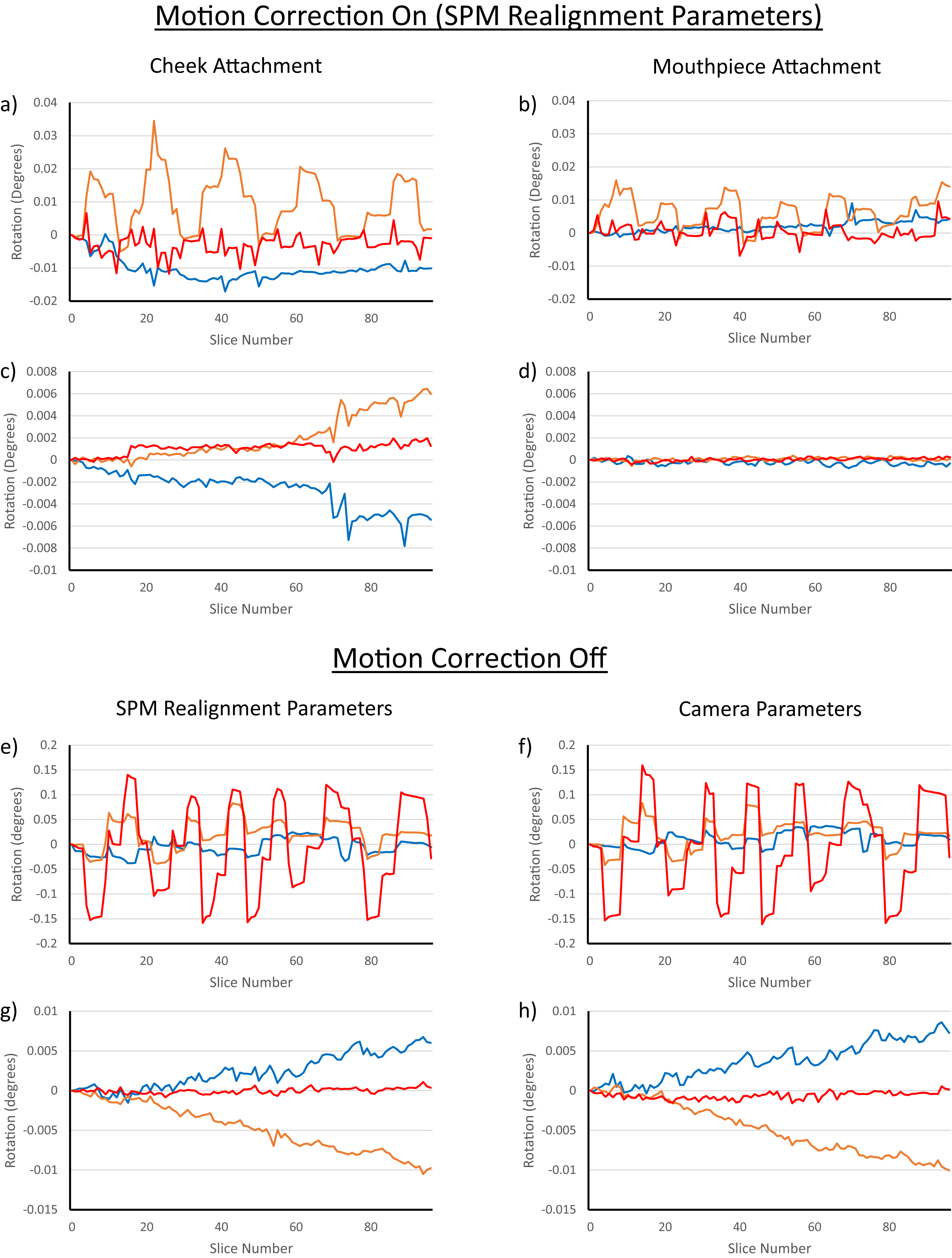
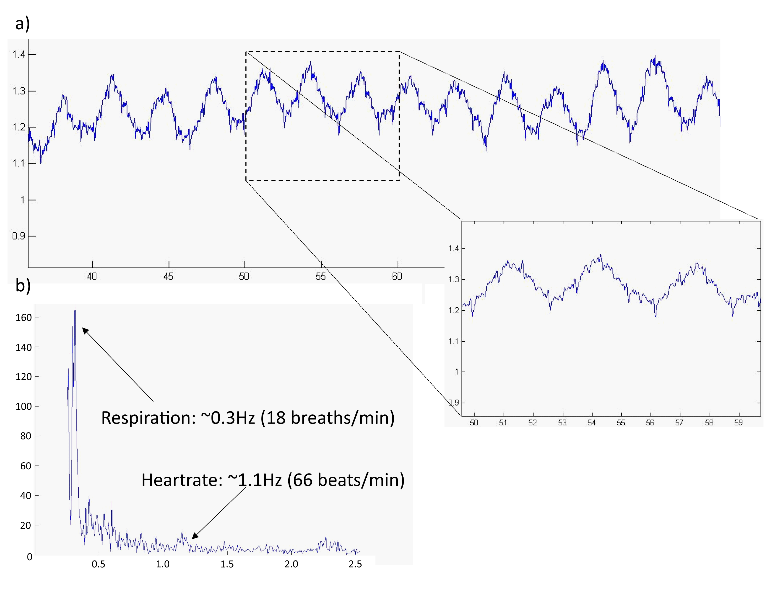
From the preliminary study, attachment to the mouthpiece showed promise in terms of adherence to motion (Figure 2) while data from cheek attachment was much worse. When no PMC was applied (Condition 2), there was strong agreement between motion parameters obtained from the camera and SPM realignment parameters. Respiration and heart rate could also be extracted from the motion data via fourier transform, attesting to the quality of the tracking (Figure 3).
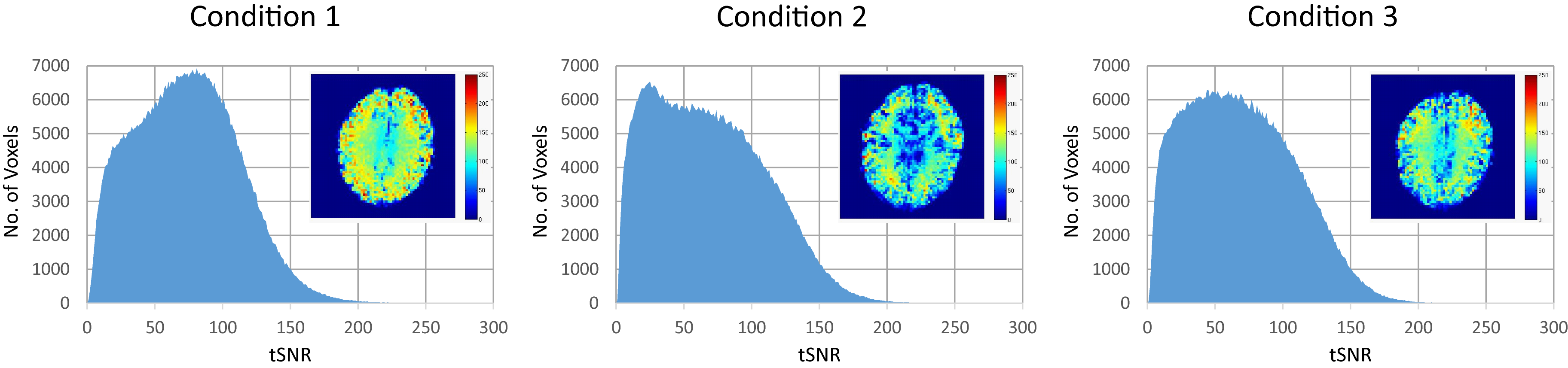
For the resting state data, tSNR was calculated for the whole brain and the results are shown in Figure 4. Application of motion correction showed a clear increase in tSNR, with Condition 1 having an average tSNR of 74, and the other two conditions both having an average tSNR of 69.
Discussion
Attachment of the marker using the mouthpiece resulted in better marker adherence. However, from both subject feedback and data analysis, the mouthpiece caused slight discomfort, resulting in increased motion and additional swallowing.
The resting state tSNR showed that the increased motion due to the mouthpiece only led to a slight reduction in data quality (Condition 2 vs Condition 3). However, when the correction by the camera is applied (Condition 1), the improvement in data quality is significant with respect to both cases with no motion correction.
Conclusion
These results testify to the effectiveness of the PMC system, providing evidence that PMC for EPI scans would improve data quality, even when only involuntary subject motion is present. However, there is the additional confounding factor of increased motion due to the presence of the mouthpiece which might affect precise voxel-to-voxel registration. Future work will test different mouthpiece designs in order to address and minimise this issue.Acknowledgements
MRI scans were funded by the Medical Research Council (MRC), UK. Pei Huang is also funded by the Agency of Science, Technology and Research (A*STAR), Singapore.References
1. Maclaren J, Herbst M, Speck O, Zaitsev M. Prospective Motion Correction in Brain Imaging?: A Review. Magn Reson Med. 2013;636:621-636. doi:10.1002/mrm.24314.
2. Stucht D, Danishad KA, Schulze P, Godenschweger F, Zaitsev M, Speck O. Highest resolution in vivo human brain MRI using prospective motion correction. PLoS One. 2015;10(7):1-17. doi:10.1371/journal.pone.0133921.
3. Todd N, Josephs O, Callaghan MF, Lutti A, Weiskopf N. Prospective motion correction of 3D echo-planar imaging data for functional MRI using optical tracking. Neuroimage. 2015;113:1-12. doi:10.1016/j.neuroimage.2015.03.013.
Figures