1273
Motion Correction for Magnetic Resonance Fingerprinting by Using Sliding-Window Reconstruction and Image Registration1School of Biomedical Engineering, Guangdong Provincial Key Laborary of Medical Image Processing, Southern Medical University, Guangzhou, People's Republic of China, 2laboratory of Biomedical Imaging and Signal Processing, The University of Hong Kong, Hong Kong SAR, People's Republic of China, 3Department of Electrical and Electronic Engineering,The University of Hong Kong, Hong Kong SAR, People's Republic of China, 4Department of Diagnostic Radiology, The University of Hong Kong, Hong Kong SAR, People's Republic of China, 5Philips Healthcare, Guangzhou, People's Republic of China, 6Department of Biomedical Engineering, Zhejiang University, Hangzhou, People's Republic of China
Synopsis
The recently proposed magnetic resonance fingerprinting (MRF) technique demonstrates to be motion insensitive, but the early motion during the acquisition can still lead to severe errors in parameter quantification. In this study, we present a novel motion correct method for MRF based on sliding-window reconstruction and image registration.
Purpose
Magnetic resonance fingerprinting (MRF)1 was reported to be motion insensitive. However, the early motion during the acquisition can still severely degrade the quality of MRF2. Motion compensated robust fingerprinting (MORF)2 utilizes combined parallel and sparse MRI techniques to reconstruct each timeframe from each spiral readout, and then correct inter-timeframe motion using image registration methods. The timeframes reconstructed from individual spiral readouts in MRF usually contain strong aliasing artifacts because of high under-sampling, and thus cannot provide sufficient information for the accurate motion estimation. Recently, the sliding-window reconstruction was shown to improve the parameter quantification accuracy in MRF3. This work aims to investigate the feasibility of correcting motion in MRF by using the sliding-window and image registration techniques.Methods
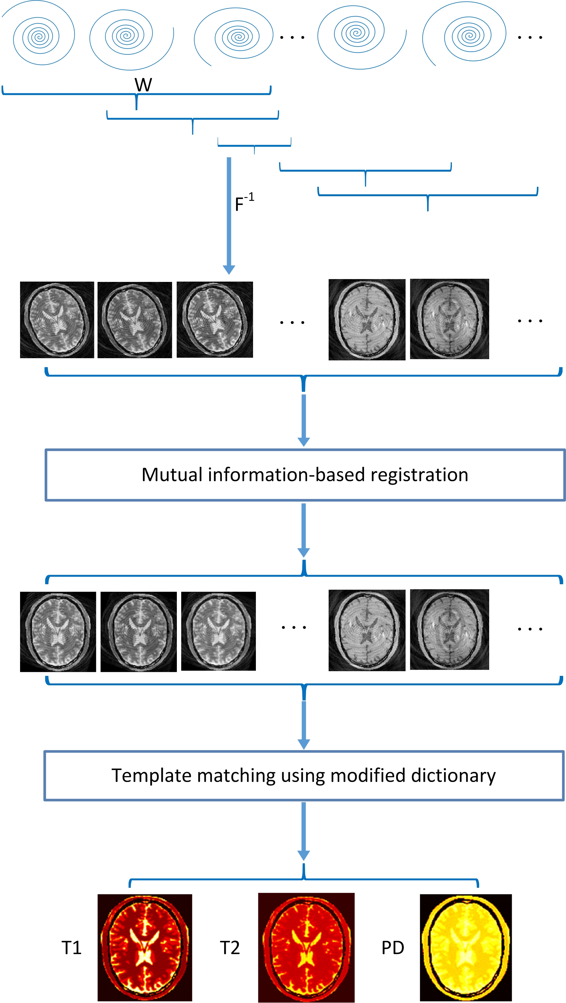
Figure 1 depicts the procedure of the proposed method. Firstly, W consecutive interleaves were chosen to reconstruct intermediate images. Secondly, the intermediate images were registered to a reference image, which was determined as the image having the strongest cross correlation with other intermediate images. Finally, the parameter maps were obtained by matching motion-corrected evolution signals to a dictionary that is adapted to the sliding-window approach3.
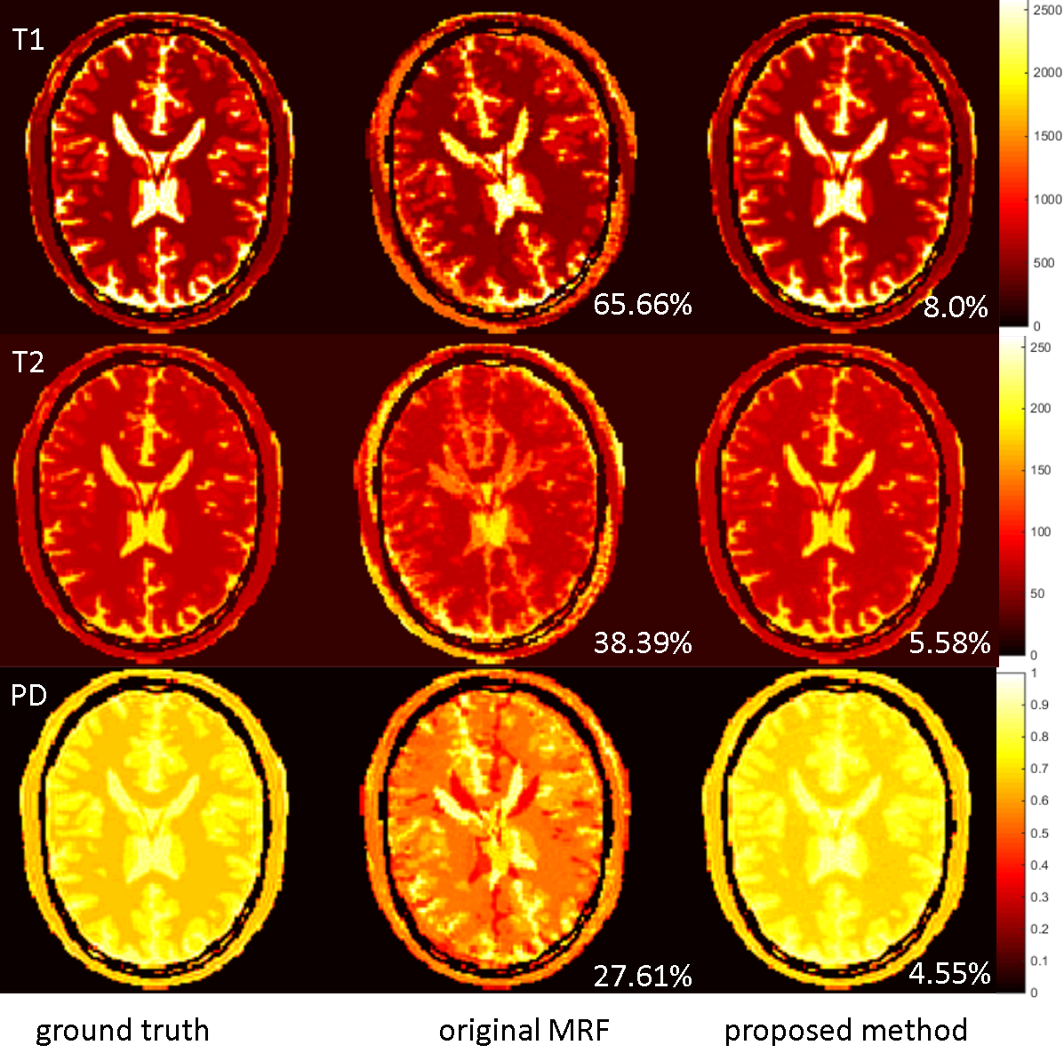
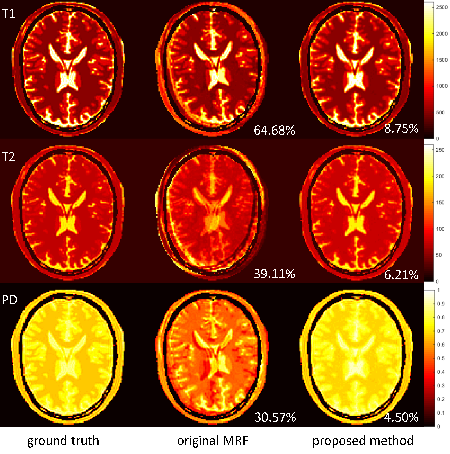
To evaluate the performance for the proposed method, simulation was implemented using a numerical brain phantom with known T1, T2, and PD maps from MNI brain atlas4. A total of 1000 brain images were generated by synthesizing the evolution signal based on MRF-FISP5 sequence with TR varying from 11.5 to 14.5ms and flip angle varying from 5 to 70 degrees. The acquisition pattern was a variable density spiral (VDS) trajectory, with the k-space center fully-sampled and the k-space edge under-sampled by a factor of 48. The spiral arm was rotated by the golden angle (222.5 degrees) after each excitation6.
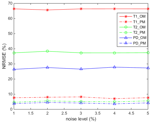
Three experiments were done. Experiment 1: rotate 20 degrees in the initial 200 timeframes, and 2% random noise was added to the 1000 complex images. Experiment 2: rotate 8 degrees, and translate 4 pixels along x direction and 3 pixels along y direction in the initial 200 timeframes; in addition, a rotation of 8 degrees, and a translation of 5 pixels along x direction and 4 pixels along y direction for timeframes from 450 to 600, and 2% noise was added. Experiment 3: a rotation of 20 degrees in the initial 200 timeframes, and the all 1000 timeframes was added with noise with varying levels from 1% to 5% with an increment of 1%. In all the three experiments, the dictionary was calculated using extending phase graph7, and the window width W was set to 12.
Results
Figures 2 and 3 show the T1, T2 and PD maps of the ground truth, original MRF and the proposed method from the experiment 1 and 2, respectively. The maps estimated by original MRF contained marked motion artifacts. In contrast, the proposed method produced maps without noticeable artifacts and closer to the ground truth. Quantitatively, the proposed motion correction method yielded substantially decreased NRMSE values than the original MRF method, in the presence of rotations and/or translations. Figure 4 shows that the proposed method consistently outperform the original MRF in terms of NRMSE (T1, T2, and PD) under varying noise levels.Discussion
The simulation results demonstrate that the proposed motion correction sliding-window approach can successfully eliminate the effect of in-plane motion on MRF. The proposed method is simple, robust and efficient, compared with the MORF method. In the current implementation, the width of the sliding-window can be further reduced by using parallel MRI techniques to reconstruct intermediate results.Conclusion
The combination sliding-window reconstruction and image registration technique can effectively correct in-plane motion in MRF.Acknowledgements
No acknowledgement found.References
1. Ma D, et al. Magnetic Resonance Fingerprinting. Nature. 2013; 495:187-193.
2. Mehta BB, et al. Image Registration and Robust Fitting for Motion Insensitive Magnetic Resonance Fingerprinting (MRF). ISMRM. 2016; p4256.
3. Cao XZ, et al. Sliding-window Reconstruction Strategy for Accelerating the Acquisition of MR Fingerprinting. ISMRM. 2016; p4200.
4. Aubert-Broche B, et al. A new improved version of the realistic digital brain phantom. Neuroimage. 2006; 32:138–145.
5. Jiang Y, et al. MR fingerprinting using fast imaging with steady state precession (FISP) with spiral readout. Magn Reson Med. 2015; 74:1621–1631.
6. Kim YC, et al. Flexible Retrospective Selection of Temporal Resolution in Real-Time Speech MRI Using a Golden-Ratio Spiral View Order. Magn Reson Med. 2011; 65:1365–1371.
7. Weigel M. Extended phase graphs: dephasing, RF pulses, and echoes-pure and simple. J Magn Reson Imaging. 2015; 41:266–295.
Figures