1255
GABA-edited echo-planar spectroscopic imaging (EPSI) with MEGA-sLASER at 7T1Danish Research Centre for Magnetic Resonance, Centre for Functional and Diagnostic Imaging and Research, Copenhagen University Hospital, Hvidovre, Denmark, 2Center for Magnetic Resonance, DTU Elektro, Technical University of Denmark, Lyngby, Denmark
Synopsis
Magnetic resonance spectroscopy (MRS) benefits from increased magnetic field-strength in terms of increased sensitivity and spectral separation and human cerebral concentrations of neurotransmitters have been measured with improved precision at 7T. We utilize the high sensitivity at 7T for accelerated magnetic resonance spectroscopic imaging (MRSI) of the gamma-aminobutyric acid (GABA) inhibitory neurotransmitter in the human brain at 7T using spectral editing (MEGA) and a semi-localized by adiabatic selective refocusing (sLASER) with echo-planar readout (EPSI). The proposed method is shown to allow for localized GABA detection and demonstrate potential for efficient imaging of GABA.
Introduction
Magnetic resonance spectroscopy (MRS) benefits from increased magnetic field-strength in terms of increased sensitivity and spectral separation of resonances. At ultra-high field-strengths (7T) using single voxel spectroscopy (SVS) techniques, human cerebral concentrations of e.g. the gamma-aminobutyric acid (GABA) inhibitory neurotransmitter has been measured with improved precision compared to lower field strengths (1). Metabolite mapping is achieved by magnetic resonance spectroscopic imaging (MRSI). The standard Chemical Shift Imaging (CSI) technique can be used for this purpose but this requires extensive acquisition time. Echo Planar Spectroscopic Imaging (EPSI) significantly accelerates MRSI by simultaneous encoding of one spatial and the temporal dimension with echo-planar gradient oscillation during signal read-out (2), and the increased sensitivity at 7T can be efficiently utilized for MRSI with EPSI. Efficient detection of human brain GABA was recently demonstrated at 7T with a SVS MEGA sequence by semi-localized by adiabatic selective refocusing (MEGA-sLASER) sequence with minimal chemical shift localization (CSL) error and with efficient spectral editing of the GABA 3.0 ppm resonance (3). The purpose was to perform MRSI of GABA in the human brain at 7T with a MEGA-sLASER spectral editing sequence using echo-planar readout.Materials and Methods
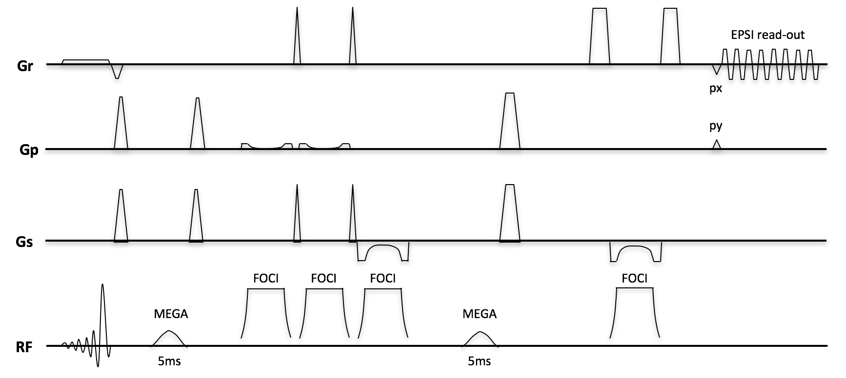
MRS measurements were performed on a healthy volunteer with a 7T MR scanner (Achieva, Philips, Cleveland, OH) interfaced to a 2-channel volume transmit head coil and 32 channel receiver array (Nova Medical, Inc., Burlington, MA). A SVS MEGA-sLASER sequence was combined with an echo-planar gradient waveform train during signal read-out (Figure 1) and was modified for optimal echo-planar gradient train onset. Gaussian pulses (5ms duration, FW95%M=100Hz) were applied at the 1.99ppm resonances (on-pulse) for refocusing of the J-coupling evolution of the 3.0 ppm GABA resonance and at the 7.0ppm (off-pulse) for the spectral editing. The pulse duration was selected to have the B0 inhomogeneity range over the imaged volume covered within the FW95%M=100Hz of the pulse. The single MRSI imaging plane was a transversal midbrain slice above the ventricles with FOV =160x160mm2 (B1=18uT, 2nd-order shimming, matrix size=16x16, in-plane voxels size=10x10mm2, slice thickness=22mm, TR=4281ms, TE=74ms, signal averages=4). The sLASER volume selection box was 80x80x22mm3 for lipid exclusion. The volume selection was obtained by an asymmetric excitation pulse combined with four adiabatic refocusing FOCI-pulses to minimize CSL-errors. The read-out bandwidth per pixel was 4kHz and the spectral bandwidth of the echo-planar gradient train was 2368Hz. The protocol consisted of one water suppressed (VAPOR, window=200Hz) scan (duration=9min) and a corresponding water-reference scan (duration=2min) with the editing pulses turned off for the latter. Spatially localized spectra were reconstructed with Matlab software (Matlab, The MathWorks, Inc.) developed in-house with a Cartesian reconstruction in the spatial and temporal dimensions and including weak temporal apodization (lw=16Hz), automatic phase corrections over coil elements and spatial locations, and eddy current compensation. The measurements were performed according to the local ethical protocols.Results and Discussion
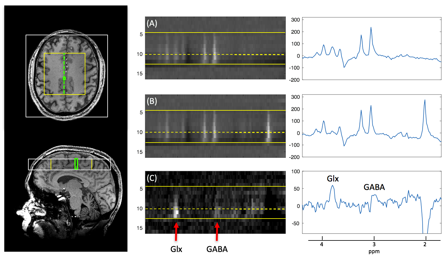
The spatially localized spectra were acquired with a signal-to-noise ratio of SNR=50 for the NAA-peak in the off-pulse acquisition, giving SNR=5 for corresponding edited GABA-peaks (Figure 2). The selected method for shimming gave a B0-variation extending a range of 50-60Hz over the sLASER selection volume, as assessed from the water reference scan. This range was well covered within the bandwidth of the Gaussian editing pulses (FW95%M=100Hz) assuring no spatial variation in the editing performance over the sLASER selection volume. Spectral frequency shifts with spatial location from B0-inhomogeneities over the sLASER selection volume were to a large extent compensated for by the water reference eddy current compensation (Figure 2, middle). The absence of positional shifts in the spatial dimension of the spectrograms of Figure 2 demonstrates minimal CSL-errors from the sLASER adiabatic refocusing. Little aliased peak disruption of the spectrums from the Cartesian reconstruction was observed in the frequency range of interest, and an optimized non-Cartesian reconstruction can further reduce the effect and related spatial blurring.Conclusion
A MEGA-sLASER sequence was combined with an optimized echo-planar read-out for accelerated MRSI while utilizing the high sensitivity at 7T field-strength. The results show in vivo localized detection of the GABA inhibitory neurotransmitter and demonstrate potential for efficient image based mapping of GABA. The technique may ultimately benefit studies of neurological and psychiatric disorders as well as studies of the healthy brain.
Acknowledgements
This work was sponsored Danish Council for Independent Research (4093-00280A, 4093-00280B and 6111-00349A).
References
1. Stephenson M, Gunner F, Napolitano A, Greenhaff PL, MacDonald IA, Saeed N, Vennart W, Francis ST, Morris P. Applications of multi-nuclear magnetic resonance spectroscopy at 7T. World J Radiol 2011; 3(4): 105-113.
2. Posse S, Tedeschi G, Risinger R, Ogg R, Le Bihan D. High speed 1H spectroscopic imaging in human brain by echo planar spatial-spectral encoding. Magn Reson Med 1995;33:34-40.
3. Andreychenko A, Boer VO, Arteaga de Castro CS, Luijten PR, Klomp DW J. Efficient Spectral Editing at 7 T: GABA Detection with MEGA-sLASER. Magn Reson Med 2012; 68:1018–1025.
Figures