1240
Thalamic functional connectivity in Multiple Sclerosis: the role of temporal thalamic sub-region in maladaptationPaola Valsasina1, Alessandro D'Ambrosio1, Milagros Hidalgo1, Elisabetta Pagani1, Bruno Colombo2, Mariaemma Rodegher2, Andrea Falini3, Giancarlo Comi2, Massimo Filippi1, and Maria Assunta Rocca1
1Neuroimaging Research Unit, San Raffaele Scientific Institute, Vita-Salute San Raffaele University, Milan, Italy, 2Department of Neurology, San Raffaele Scientific Institute, Vita-Salute San Raffaele University, Milan, Italy, 3Department of Neuroradiology, San Raffaele Scientific Institute, Vita-Salute San Raffaele University, Milan, Italy
Synopsis
We compared resting state (RS) functional connectivity (FC) of five thalamic sub-regions (frontal, motor, post-central, occipital and temporal) between patients with multiple sclerosis (MS) and healthy controls. There was an overall increase of intra- and inter-thalamic RS FC for almost all thalamic sub-regions, apart from the temporal thalamic sub-region, which showed reduced intra-thalamic RS FC and higher RS FC with the fronto-parietal somatomotor cortex. Compared to cognitively preserved, cognitively impaired MS patients had lower RS FC between thalamic sub-regions and caudate nucleus, anterior cingulate cortex, as well as higher RS FC between thalamic sub-regions and several temporal areas.
Purpose
In spite of the well-known importance of the thalamus in multiple sclerosis (MS), only limited data on whole and sub-regional thalamic functional connectivity (FC) are available [1]. Aim of this study was to investigate the sub-regional thalamic resting state (RS) FC and its relationship with clinical and cognitive measures in multiple sclerosis (MS) patients.Methods
Diffusion tensor (DT), 3D T1-weighted and RS functional MRI data were acquired from 187 right–handed MS patients and 94 age- and sex-matched healthy controls (HC). MS patients underwent a clinical assessment, with rating of the EDSS score [2], and a cognitive evaluation [3]. Patients were considered cognitively impaired if they had abnormal scores in at least two cognitive tests [4]. DT MRI data were used to parcellate the thalamus into five sub-regions, according to their structural connectivity profile (frontal, motor, post-central, occipital and temporal, Figure 1), as described in details elsewhere [5]. For each sub-region, a seed-based RS FC analysis was performed. Multiple regression models were run to assess correlations between thalamic RS FC and clinical/cognitive variables.Results
Compared to HC, MS patients had higher intra- and inter-thalamic RS FC in almost all thalamic sub-regions. Conversely, temporal thalamic sub-region showed reduced intra-thalamic RS FC and higher RS FC with the fronto-parietal somatomotor cortex (Figure 2). Compared to cognitively preserved, cognitively impaired MS patients had lower RS FC between thalamic sub-regions and caudate nucleus, anterior cingulate cortex, as well as higher RS FC between thalamic sub-regions and several temporal areas (Figure 3). This latter finding was correlated with poor motor and cognitive performance. Lower RS FC between thalamic sub-regions and caudate and cingulate cortex was correlated with worst motor/cognitive performance.Conclusions
In MS, the different behavior of temporal thalamic sub-region, compared to other thalamic sub-regions, could contribute to explain the variability of thalamic RS FC findings in previous studies. The increased RS FC between temporal thalamic sub-region and temporal/frontal regions seen in MS patients compared to HC, as well as in cognitively impaired compared to cognitively preserved MS patients is likely to be a maladaptive mechanism, associated with clinical/cognitive impairment.Acknowledgements
This study has been partially supported by a grant from FISM 2011/R/19 and Italian Ministry of Health (GR-2009-1529671).References
[1] Schoonheim M., et al. Neurology 2015; 84:776-83. [2] Kurtzke J.F. Neurology 1983; 33:1444-52. [3] Rao S., et al., Neurology 1991. [4] Amato M.P., et al. Mult Scler 2006; 12:787-93. [5] Bisecco A., et al. Hum Brain Mapp 2015; 36:2809-25.Figures
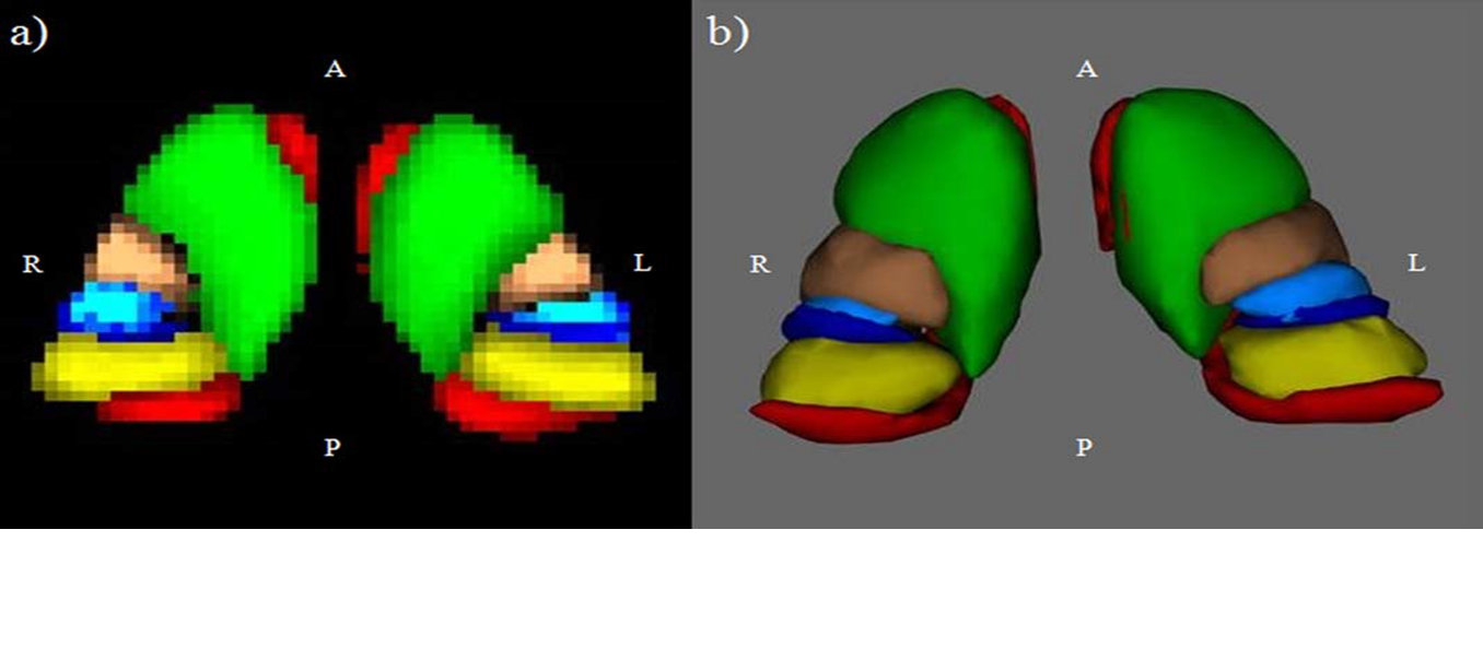
Thalamic connectivity defined regions (CDRs)
probability maps from HCs, thresholded at 33%, in Montreal Neurological
Institute space: (a) axial 2D view; (b) ventral 3D view. Green=frontal CDR,
copper=motor CDR, blue=postcentral CDR, red=temporal CDR, yellow=occipital CDR.
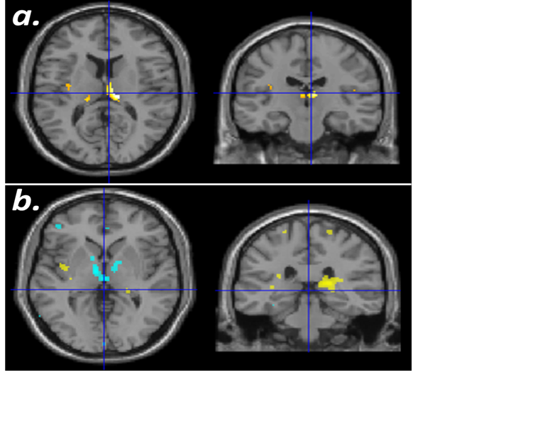
Sub-regional thalamic RS FC in MS patients
compared to HC.
(a) The frontal-thalamic sub-region of MS
patients showed, as the majority of thalamic sub-regions, higher intra and
inter thalamic RS FC (yellow).
(b) The temporal-thalamic sub-region of MS
patients showed lower intra-thalamic RS FC (cyan) and higher RS FC with frontal
and hippocampal areas (yellow).
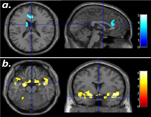
(a) The
frontal-thalamic sub-region of cognitively impaired (CI) MS patients showed, as
almost all thalamic sub-regions, lower RS FC with caudate nucleus and anterior
cingulate cortex (cyan).
(b.) The
temporal-thalamic sub-region of CI MS patients showed higher RS FC with
inferior frontal areas and para-hippocampal gyrus (yellow).