1226
Universal Size-Optimized 48-Channel Phased-Array Receive Head Coil for 3T Clinical fMRI/Silent Imaging Application1Engineering, GE Healthcare Coils, Aurora, OH, United States, 2Engineering, GE Healthcare, Waukesha, WI, United States
Synopsis
Typical high
density phased-array head coils are design to fit tightly around the head to achieve increasing SNR and improving
acceleration limiting space and excluding its use for larger patients. This
study shows the results for an optimized 48-Channel phased-array receive head coil
design on a universally sized football helmet-shaped former (fitting to more
than 99th - percentile US male) providing similar SNR and improved
acceleration compared to a 32-Channel close-fitting design coil. The
performance of this array is demonstrated in highly accelerated head images on
variously sized human subjects. This coil is optimized for Hyperband, fMRI and
Silent imaging.
Introduction
Close-fitting designs are used in high density phased-array head coils to achieve increased SNR and improved acceleration [2]. Some patient populations do not fit in this type of close-fitting design coil. This also decreases patient comfort. In this paper, a novel high field universal size-optimized 48-channel phased-array receive head coil for fMRI/Silent Imaging on a 3.0 T whole body MRI system is presented. A prototype of the 48-channel phased-array head coil was constructed on a universally sized former (fitting more than 99th - percentile US male). The SNR and acceleration performances were quantitatively evaluated in phantom and human imaging and compared to a clinical 32-channel phased array built on a close-fitting shaped former (fitting to 5th- percentile US male). The performance of this coil was also compared to 21-channel clinical Head-Neck Array (HNA). The performance of the 48-channel head coil is demonstrated in highly accelerated diffusion-weighted imaging, functional MRI (fMRI) and Silent imaging.Methods
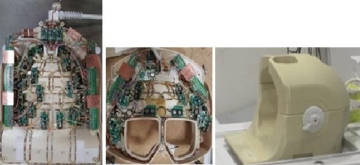
A universally sized football helmet-shaped former was developed using SolidWorks™ 3-D modeling. A range of head sizes were evaluated, allowing for a 15mm foam pad beneath the head and 1 cm spacing above the forehead for large head size. Optimal major and minor axes were found to be 230 mm A-P and 230 mm L-R, respectively, for the inner surface. A split between top and bottom sections of the former was placed 45 mm above the center to accommodate more elements in the bottom section. Full brain coverage was achieved by sixteen of the elements located in the top former and twenty-eight more elements were placed in the bottom former in the same z-location, as indicated in Fig. 1. Four more elements then added to the bottom array to provide coverage of the extended C4. Five tiles of posterior coil elements and four tiles of anterior coil elements are placed in 24cm FOV to increase the acceleration factor. These tiles were staggered in the RL direction. This head coil was tuned to 127.73MHz. Nearest neighboring elements were decoupled by overlapping and next nearest neighbor and more distant elements were decoupled by preamplifiers. Coil is decoupled from body coil using the custom MEMS (Micro Electro Mechanical System) switch [4] and optimized to reduce the RF switching time for Silent imaging. The B1 sensitivity map and g-factors for R=2, 3, 4 and 5 were calculated using the finite difference time domain (FDTD) method for the fields on 21-channel HNA, 32-channel close-fitting coil and 48-channel head coil. The SNR and acceleration performances were measured in phantom and human imaging. In vivo volunteer imaging using the optimized 48-channel phased array receive coil at 3.0T incorporating Hyperband and ASSET sequences for fMRI was performed for reduction factors of R=2, 3 and 4. The coil was developed and tested on a 128-channel 3T GE scanner (Signa Architect, GE Healthcare, Waukesha, USA).Results
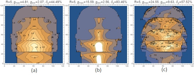
SNR maps using optimal reconstruction are shown in Fig. 2. For SNR comparison, Spin Echo images were obtained in phantom and human subjects. At the central region, the 48-channel coil shows 4% better SNR than 32-channel close-fitting coil and 15% better SNR than 21-channel HNA on a loading shell phantom. At the entire image region, the 48-channel head coil shows 8% lower SNR than the 32-channel close-fitting coil and 23% better SNR than 21-channel HNA on a loading shell phantom. On average, at the central region, the 48-channel head coil shows ~10.5% better SNR than 32-channel close-fitting coil and ~13.5 % better SNR than 21-channel HNA on two volunteers. On average, at the entire image region, the 48-channel head coil shows slightly lower SNR (~1%) than the 32-channel close-fitting coil and ~28% better SNR than 21-channel HNA on two volunteers. Figure 3 shows the maximum g-factor results at R=5 for the three coils for 1D acceleration in the SI coronal. Table 1 summarizes the maximum g-factor at 2, 3, 4 and 5 for 1D acceleration in the SI coronal. The benefits of increased channel number for reducing g-factor are clear. Figure 4 shows in vivo volunteer images using the optimized 48-channel head coil.Discussion
The simulation results of B1 sensitivity maps and g-factors are matched to the results of the loading shell phantom and in vivo images.Conclusions
The universal size-optimized 48-channel head coil is an open design that accommodate large heads. The sagittal view shows good whole-brain coverage, and lower g-factors in the axial, sagittal and coronal planes compared to standard 32-channel close-fitting coil and 21-channel HNA. The SNR performance is similar or better compared to 32-channel close-fitting design coil and 21-channel HNA.
Acknowledgements
No acknowledgement found.References
1. Roemer, P.B., et al., The NMR phased array. MRM, 1990; 16(2):192-225.
2. Wiggins, G.C., et al., 96-Channel Receive-Only Head Coil for 3 Tesla: Design Optimization and Evaluation. MRM, 2009; 62:754–762.
3. Pruessmann K.P., et al., SENSE: sensitivity encoding for fast MRI. MRM, 1999;42:952–962.
4. Spence, D., et al., Custom MEMS Switch for MR Surface Coil decoupling. ISMRM, 2015:0704.
Figures