1223
In-vivo (8x4) 32-ch Tx-only Body Array for UHF MRI.1Bioengineering, University of Pittsburgh, Pittsburgh, PA, United States, 2Radiology, University of PIttsburgh, Pittsburgh, PA, United States, 3Bioengineering, University of Pittsburgh, 4MR Research & Collaboration, Siemens Healthineers, NY, 5Bioengineering, University of Pittsburgh, PA, 6Radiology, University of PIttsburgh
Synopsis
The Clinical and Research potential of MRI especially for Whole Body imaging is limitless. Body MR exams are growing part of total clinical MRI exams today. It however faces considerable challenges such as significant rise in RF power deposition in tissue and daunting high field inhomogeneities/signal voids across the anatomy of interest most especially at 7T and higher. This study aims at utilizing the intrinsic sensitivity advantage of 7T by exploring a 32-ch transmit coil design in order to generate a circularly polarized field with homogeneous and extended coverage in abdominal/body regions (liver, kidney, and abdomen in general) at 7T.
Target Audience: Researchers who are interested in Ultra-high Field (UHF) Body imaging
Background: The clinical and research potential of MRI especially for Whole Body imaging is limitless (1). When compared to commercially available scanners in the market today (<=3T), high field imaging provides almost twice the signal to noise ratio (SNR), high spatial-temporal resolution, and reduced scan time (2). Body MR exams are growing part of total clinical MRI exams today (3). It however faces considerable challenges such as significant rise in RF power deposition in tissue and daunting high field inhomogeneities/signal voids across the anatomy of interest most especially at 7T and higher (4, 5). This study aims at utilizing the intrinsic sensitivity advantage of 7T by exploring a 32-ch transmit coil design in order to generate a circularly polarized field with homogenous and extended coverage in abdominal/body regions (liver, kidney, and abdomen in general) at 7T.
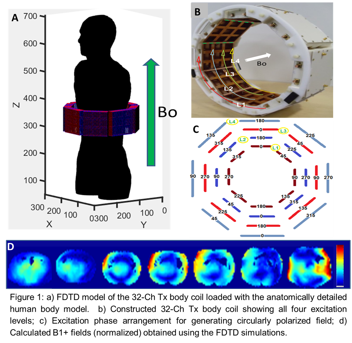
Methods: A 32-channel transmit body array contains 8 decoupled sets/sides of highly coupled 4-channel Tic Tack Toe arrays(6). Each side is placed in an octagon arrangement as shown in Fig. 1 (A, B). Each side has 2x2 coaxial elements. Each panel has 4 independent Tx channels as shown in Fig. 1 (B). While the RF field distributions from each side are highly coupled, each isolated panel creates RF field distributions that are highly decoupled from the other sides. The finite difference time domain (FDTD) method was used to numerically simulate the coil and human model as single system in order to create the B1+ field distributions. Fig. 1 (A) shows FDTD model/mesh of the coil and body/torso. All the experiments were conducted with the 7T Siemens Magnetom scanner. In-vivo imaging data were collected on three healthy volunteers under University approved Institutional Review Board (IRB). This study was conducted on parallel transmit (pTx) system. An eight pTx channels were connected to eight 1:4 Wilkinson power divider and constant phase shifters in order to produce a pseudo circularly polarized (CP) mode. There are four Z levels (L) in the coil as shown in Fig. 1 (C). Looking into the B0 direction, the phases of the voltages were rotating clockwise with increments of 45° on each level with 180o phase difference between different levels shown in Fig. 1(C). Sequence parameters are given as follows: B1+ field mapping: Slice Thickness: 3 mm, TR/TE: 13740/3.13 ms, No. of Avgs: 1, No. of phase encoding steps: 96 , Pixel bandwidth: 491, TA: 00.56 min, Fov: 300x300 , Resolution: 0.32 pixels per mm; ii) T1 VIBE: Slice Thickness: 3 mm, TR/TE: 5/1.48 ms, No. of Avgs: 1, No. of phase encoding steps: 146, Pixel bandwidth: 488, FA: 27, TA: 00.36 min, Fov: 226x330 , Resolution: 0.77 pixels per mm, Voxel size: 1.29x1.29x3.00 mm
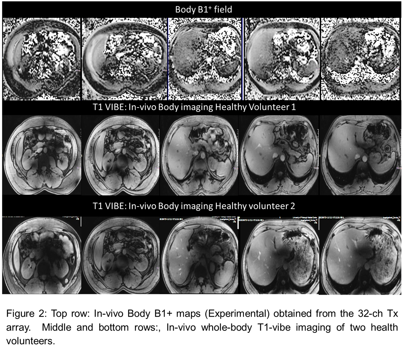
Results: All the 32 elements were easily tuned and matched to 50 ohms (< -18 dB). When loaded with different human subjects, these scattering matrix values change very insignificantly due to inherent highly coupled behavior. Fig. 1 (D) shows numerically simulated B1+ field (at ~300 MHz) in anatomically detailed model of the human body/torso. Fig. 2 (top row) shows experimentally mapped B1+ field from a healthy volunteer. Two healthy volunteer were scanned using T1VIBE sequence as shown in Fig 2 (middle and bottom rows). The simulations and experimental data show promising results in the liver (portal vein and its branches, inferior vena cava and it’s branching veins), and kidney (first and second order renal artery) of human body, overcoming the severe RF penetration issues and undesired signal voids normally encountered with body imaging at UHF (7T).
Discussion and Ongoing work: In spite of B1+ field inhomogeneity challenges, the proposed coil design would be highly beneficial in in attaining homogenous abdominal imaging. This study was carried out using a 32-ch transmit only RF array (with a parametric coupling design approach) without receive-only insert, thus SNR/contrast was compromised. Future will boost signal/contrast-noise ratio using multichannel receive-only array in conjunction with the transmit coil design. Thus utilizing 32 ch TTT Body coil at UHF shows excellent potential of whole body imaging at ultra-high field (7T) with acceptable SAR limits.
Acknowledgements
No acknowledgement found.References
1. Kraff O, Fischer A, Nagel AM, Monninghoff C, Ladd ME. MRI at 7Tesla and above: demonstrated and potential capabilities. . JMR. 2015;41:13-33.
2. Vaughan JT, Garwood M, Collins CM, et al. 7T vs. 4T: RF power, homogeneity, and signal-to-noise comparison in head images. Magnetic Resonance in Medicine. 2001;46(1):24-30. doi: Doi 10.1002/Mrm.1156. PubMed PMID: WOS:000169561000005.
3. Smith-Bindman R, Miglioretti DL, Larson EB. Rising Use Of Diagnostic Medical Imaging In A Large Integrated Health System. Health Affairs. 2008;27(6):1491-502. doi: DOI 10.1377/hlthaff.27.6.1491. PubMed PMID: WOS:000260769300005.
4. de Moortele V, Akgun C, Adriany G, Moeller S, Ritter J, Collins CM, Smith MB, J. V, Ugurbil K. B1 destructive interferences and spatial phase patterns at 7 T with a head transceiver array coil. MRM. 2005;54(6):1503-18.
5. Vaughan TT, Snyder CJ, DelaBarre LJ, Bolan PJ, Tian J, Bolinger L, Adriany G, Andersen P, Strupp J, Ugurbil K. Whole-Body Imaging at 7T: Preliminary Results. Magnetic Resonance in Medicine. 2009;61(1):244-8. doi: Doi 10.1002/Mrm.21751. PubMed PMID: WOS:000261914000030.
6. Kim J, Krishnamurthy N, Santini T, Zhao Y, Zhao T, Bae KT, Ibrahim TS. Experimental and numerical analysis of B1(+) field and SAR with a new transmit array design for 7T breast MRI. JMR. 2016;269:55-64.
Figures