1184
Regional Detection of Lung Injury using Hyperpolarized Xenon-129 Mapping of Blood Hematocrit in a Rat Model Involving Partial-Lung Irradiation1Peter Gilgan Centre for Research and Learning, The Hospital for Sick Children, Toronto, ON, Canada, 2Department of Medical Biophysics, University of Toronto, Toronto, ON, Canada, 3Radiation Medicine Program, Princess Margaret Cancer Centre, Toronto, ON, Canada, 4Department of Radiation Oncology, University of Toronto, Toronto, Canada
Synopsis
Dissolved 129Xe imaging holds promise for the detection of early functional decline due to radiation-induced lung injury through the extraction of regional quantitative parameters relating to lung physiology and gas exchange. In this work a spiral-IDEAL imaging technique was used with a partial-lung irradiation rat model to investigate regional changes lung function associated with injury and compare to histology. A significant reduction in capillary hematocrit (HCT) was observed in the vicinity of irradiation. These results were in agreement with quantitative histology of red blood cells. Imaging results were more sensitive than whole-lung spectroscopy, which was performed simultaneously.
Purpose
MRI of hyperpolarized (HP) 129Xe dissolved in the parenchymal tissue and red blood cells (RBCs) of the lungs enables the investigation of gas exchange between these compartments using chemical shift saturation recovery (CSSR) at varying saturation delay times1,2. CSSR has shown promise for early detection of radiation-induced lung injury (RILI) in previous animal studies3,4. However, these studies involved whole-thorax irradiation models and spectroscopic analysis, thereby obscuring any regional differences. Regional differences in gas exchange due to hemithoracic irradiation have been demonstrated using a spiral-IDEAL imaging approach5, but these differences were observed between left and right lungs only and were not correlated with histology of RBCs. Additionally, whole or hemithoracic irradiation may introduce cardiac damage, confounding RBC measurements. In this study we performed spiral-IDEAL HP 129Xe imaging of a rat model of RILI involving partial-lung irradiation, resulting in regional differences in injury avoiding the heart. The results of this work are correlated with regional analysis of relative RBC volume determined by histology.Methods
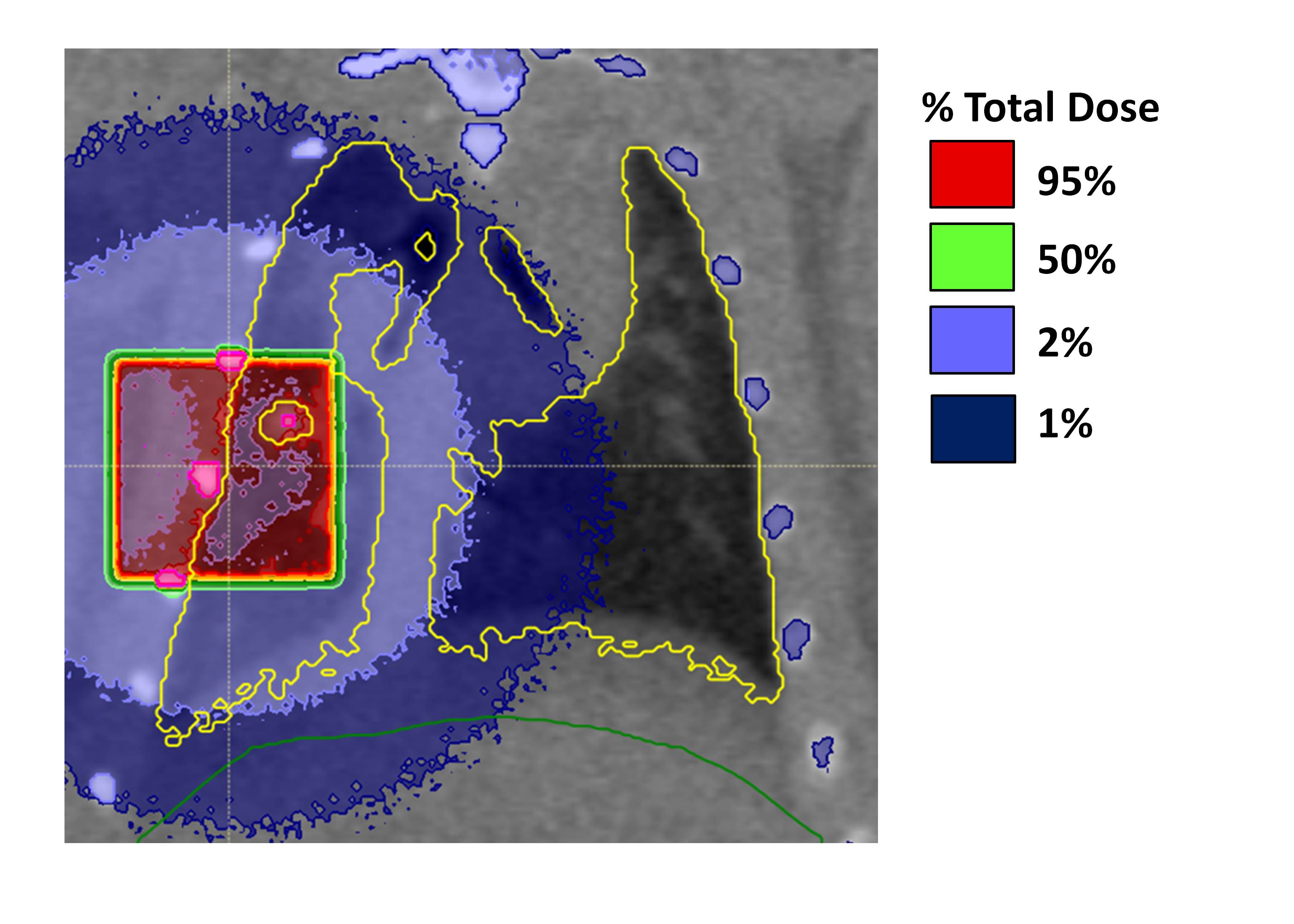
Two cohorts (N=6 each) of Sprague-Dawley rats were used. One underwent partial-lung irradiation using an image-guided small animal irradiator (X-Rad 225Cx, PXi, North Brantford, CT) with the second serving as controls. A single dose of 20Gy photons irradiated the right medial lung in the coronal direction, avoiding the heart (Fig. 1).
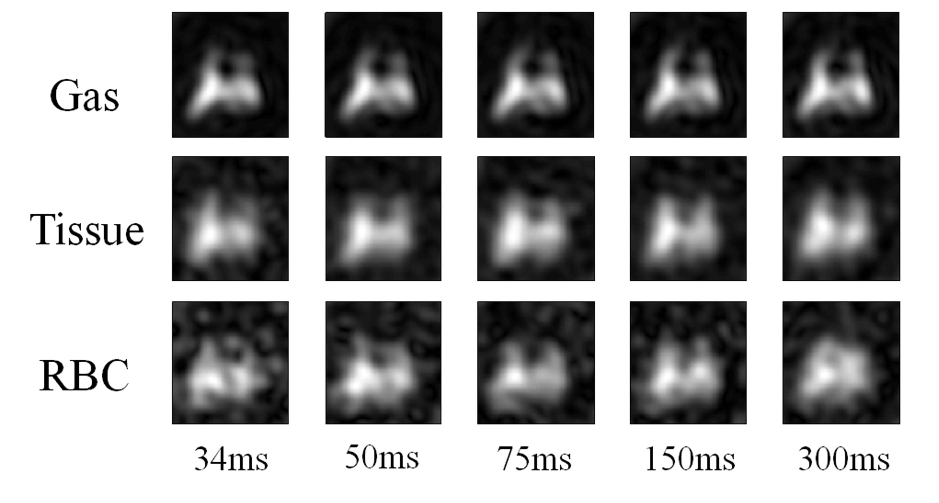
Imaging was performed 4 weeks post-irradiation on a 1.5T MRI (Signa HDxt, GEHC, Waukesha, WI) with a birdcage rat coil (Morris Instruments, Ottawa, ON). Rats were ventilated as previously described3. A 4-point spiral-IDEAL6,7 sequence was used (FOV=5×5cm2, Cartesian Matrix=16×16, TE=0.3ms, ΔTE=700μs). An additional acquisition was performed with imaging gradients nulled for spectroscopy. Coronal projections were acquired at five saturation delay times by varying the repetition time (TR) between acquisitions (34ms, 50ms, 75ms, 150ms, 300ms) as shown in Fig. 2. Multiple averages were acquired to ensure similar SNR across all timepoints. Isotopically enriched (~86%) 129Xe was hyperpolarized using a commercial polarizer (Model 9800, Polarean, Durham, NC) yielding polarizations of ~15%.
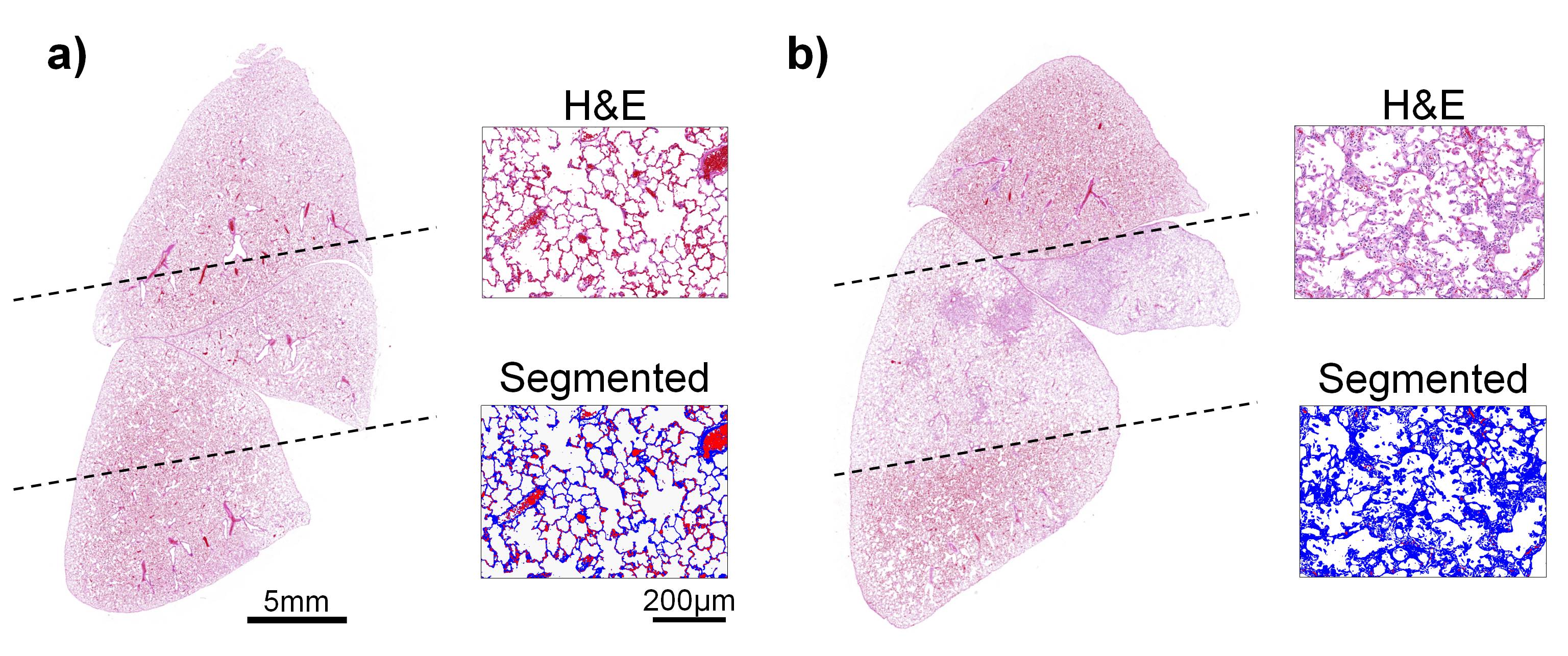
Following imaging, rats were euthanized and the lungs were removed with major vessels ligated, to retain blood. After fixation, 5μm sections from the middle of the coronal plane were H&E stained. Twenty-five images from the right medial lung were captured and segmented into three clusters (airspace, tissue, RBC)8. The percent RBC area (PRA) of the irradiated region was calculated as the number of RBC pixels relative to the total FOV9, determining relative RBC volume.
Reconstruction and analysis were performed in MATLAB (MathWorks, Natick, MA). Images and spectra were scaled and normalized by the gas-phase as described previously2,10. Tissue and RBC spiral-IDEAL images were simultaneously fit to the Model of Xenon Exchange (MOXE) and maps of hematocrit (HCT) were calculated2. Statistical analysis was performed in GraphPad Prism (San Diego, CA). Two-tailed t-tests characterized the difference between means. Two-tailed Pearson correlation with a 95% confidence interval was used to compare imaging to histology.
Results
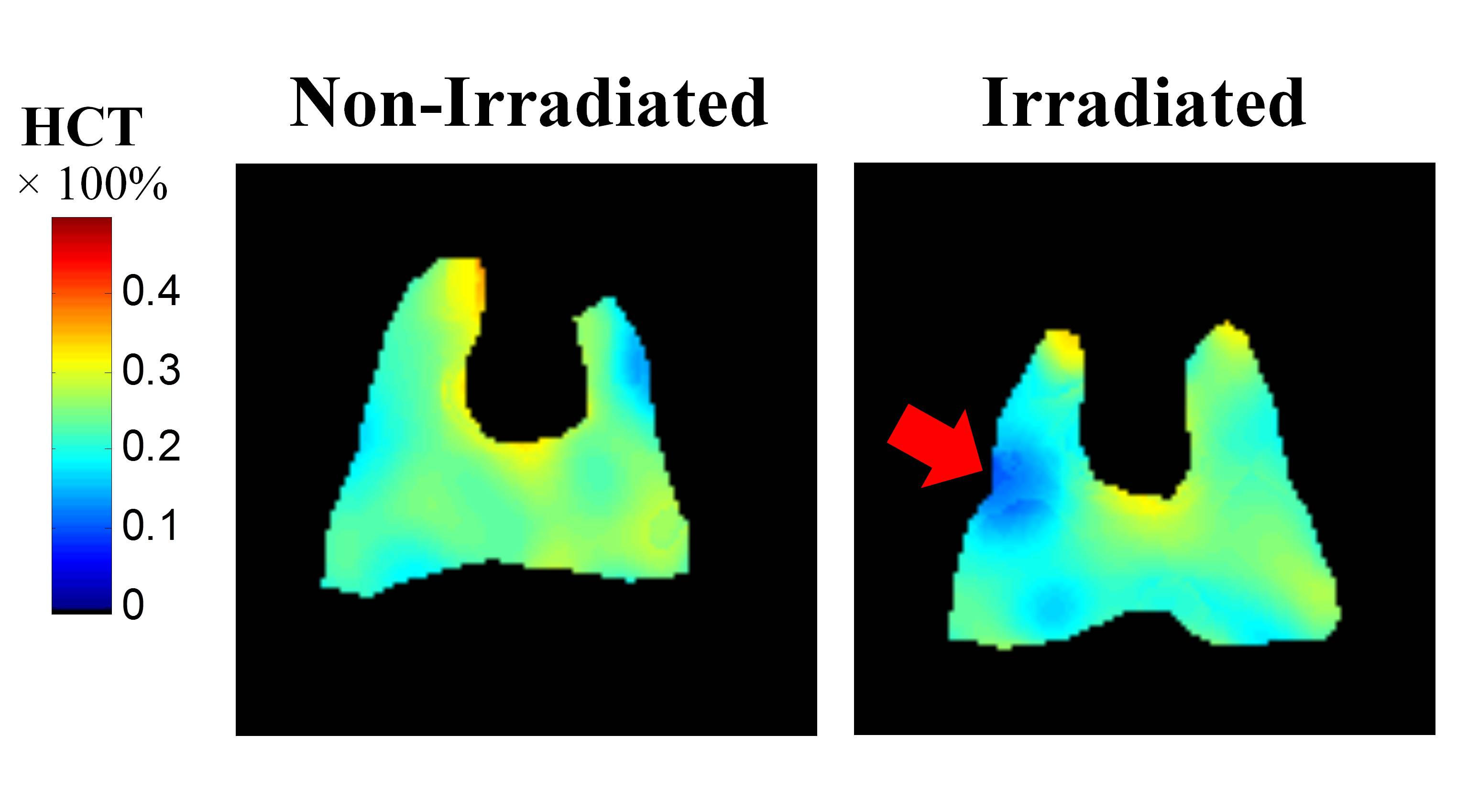
Representative HCT maps are shown
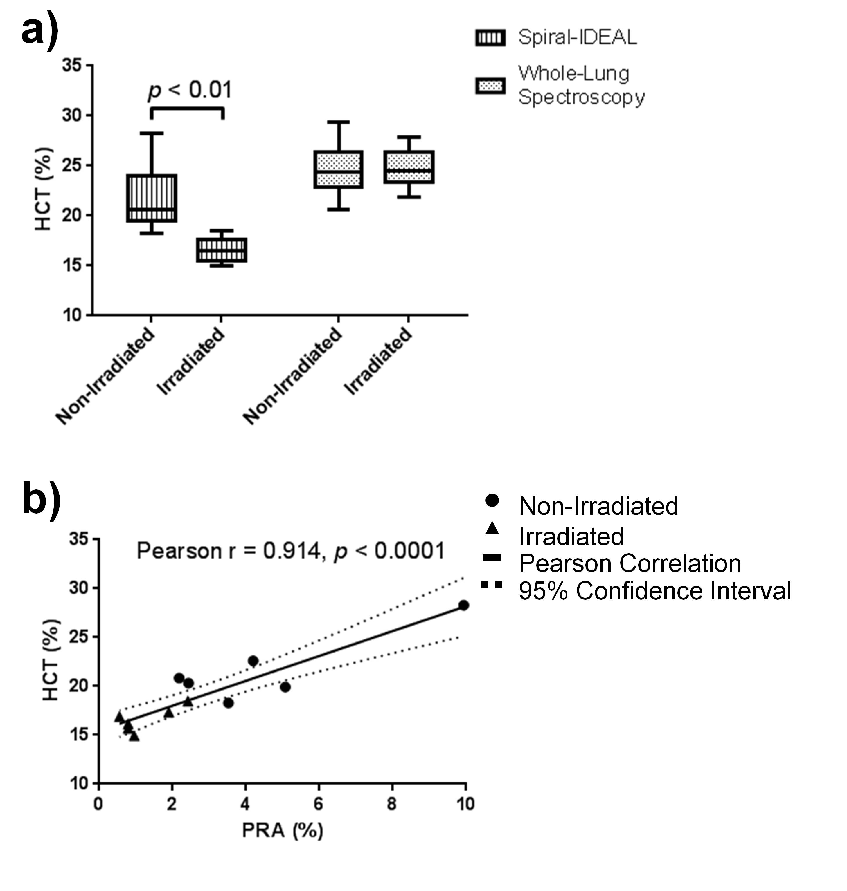
in Fig. 3. MRI results are shown in Fig. 4a. ROI analysis of the right medial
lung yielded a significant decrease (p<0.01)
in HCT in the irradiated rats compared to non-irradiated rats (16.6%±1.3% and 21.7%±4.0%,
respectively). HCT values calculated from whole-lung CSSR were not
significantly different between irradiated and non-irradiated rats (24.7%±2.0% and 24.6%±2.9%,
respectively). Imaging results were consistent with histologically-observed
decreases in blood (Fig. 5). PRA was significantly decreased (p<0.05) in the irradiated cohort
compared to the non-irradiated cohort (1.23%±0.76%
and 4.55%±2.83%, respectively). A strong positive correlation
between HCT and PRA was observed (Fig. 4b).
Discussion
HCT measured with spiral-IDEAL was significantly decreased in the region of irradiation in agreement with histologically determined PRA. This work illustrates that for focal injury, regional detection is not only desirable, but necessary since whole-lung spectroscopy HCT was not significantly different, suggesting the effect of damage may be obscured by partial-volume effects. The physiological source of this RBC reduction requires further investigation. A reduction of vessel density through vessel destruction and/or shunting of perfusion away from the injured region may be the cause. In future, the imaging described in this work may be used to answer radiobiological questions about damage to lung vasculature. This method holds additional promise for RILI detection through other MOXE parameters that are not discussed here (e.g. septal thickness, capillary transfer time). Clinical implementation of spiral-IDEAL may allow for early RILI detection during radiotherapy, potentially allowing for intervention (e.g. treatment plan adjustment, administration of pharmaceuticals) improving treatment outcomes.Conclusion
Spiral-IDEAL imaging is sensitive to regional HCT reductions associated with irradiation in a rat model, in agreement with histology.Acknowledgements
This work was supported by a NSERC, CIHR, and RESTRACOMP. Thanks to Dr. Richard Hill and Dr. Ozkan Doganay for assistance with the irradiation scheme and MRI methodology, respectively. The authors would like to recognize the Spatio-Temporal Targeting and Amplification of Radiation (STTARR) facility for assistance with irradiations. Special thanks to Nikhil Kanhere, Andras Lindenmaier, Yonni Friedlander, and Felipe Morgado for assistance with imaging experiments.References
1. Ruppert K et al. NMR of hyperpolarized 129Xe in the canine chest: Spectral dynamics during a breath-hold. NMR in Biomedicine. 2000;13(4):220–228.
2. Chang Y V. MOXE: A model of gas exchange for hyperpolarized 129Xe magnetic resonance of the lung. Magnetic Resonance in Medicine. 2013;69(3):884–890.
3. Fox MS et al. Detection of radiation induced lung injury in rats using dynamic hyperpolarized 129Xe magnetic resonance spectroscopy. Medical Physics. 2014;41(7):72302.
4. Li H et al. Quantitative evaluation of radiation-induced lung injury with hyperpolarized xenon magnetic resonance. Magnetic Resonance in Medicine. 2016;76(2):408–416.
5. Doganay O et al. Quantification of regional early stage gas exchange changes using hyperpolarized 129Xe MRI in a rat model of radiation-induced lung injury. Medical Physics. 2016;43(5):2410–2420.
6. Wiesinger F et al. IDEAL spiral CSI for dynamic metabolic MR imaging of hyperpolarized [1- 13C]pyruvate. Magnetic Resonance in Medicine. 2012;68(1):8–16.
7. Doganay O et al. Hyperpolarized 129 Xe imaging of the rat lung using spiral IDEAL. Magnetic Resonance in Medicine. 2015;76(2):566–576.
8. Sieren JC et al. An automated segmentation approach for highlighting the histological complexity of human lung cancer. Annals of Biomedical Engineering. 2010;38(12):3581–3591.
9. Takahashi A et al. Noninvasive assessment for acute allograft rejection in a rat lung transplantation model. Physiological Reports. 2014;2(12):e12244–e12244.
10. Chang Y V. et al. Quantification of human lung structure and physiology using hyperpolarized 129Xe. Magnetic Resonance in Medicine. 2014;71(1):339–344.
Figures