1157
Brain regions associated with reward induced by optogenetic stimulation at the medial prefrontal cortex1National Institute on Drug Abuse (NIDA-IRP), NIH, BALTIMORE, MD, United States
Synopsis
Optogenetics and fMRI were combined to examine the neural activity underlying the rewarding behavior mediated by the medial prefrontal cortex (MPFC) in rats. Animals were trained to press lever for optogenetic self-administration in MPFC. FMRI showed that MPFC photostimulation activated many regions known to receive MPFC afferents. Notably, the activation of hypothalamus, agranular insula, and ventral striatum was positively correlated with the lever press. Our finding may shed light on brain circuits involved in therapeutic effects of recent deep brain stimulation studies in major depression, in which the MPFC plays an important role.
Introduction
The medial prefrontal cortex (MPFC) plays an important role in mood regulation. Deep brain stimulation (DBS) delivered at the MPFC has been found to have treatment efficacy in major depression.1, 2 Preclinical research suggests an involvement of this region in positive emotion or reward. For example, rats learn to self-stimulate with high-frequency electricity delivered at the MPFC.3-5 In the present study, we incorporated optogenetic stimulation and whole brain fMRI to provide a more complete view of brain circuits underlying positive emotional effects induced by MPFC stimulation.Methods
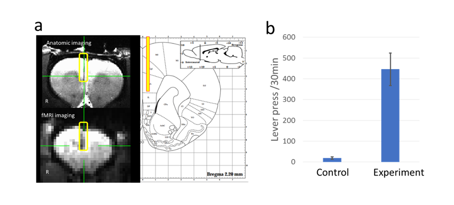
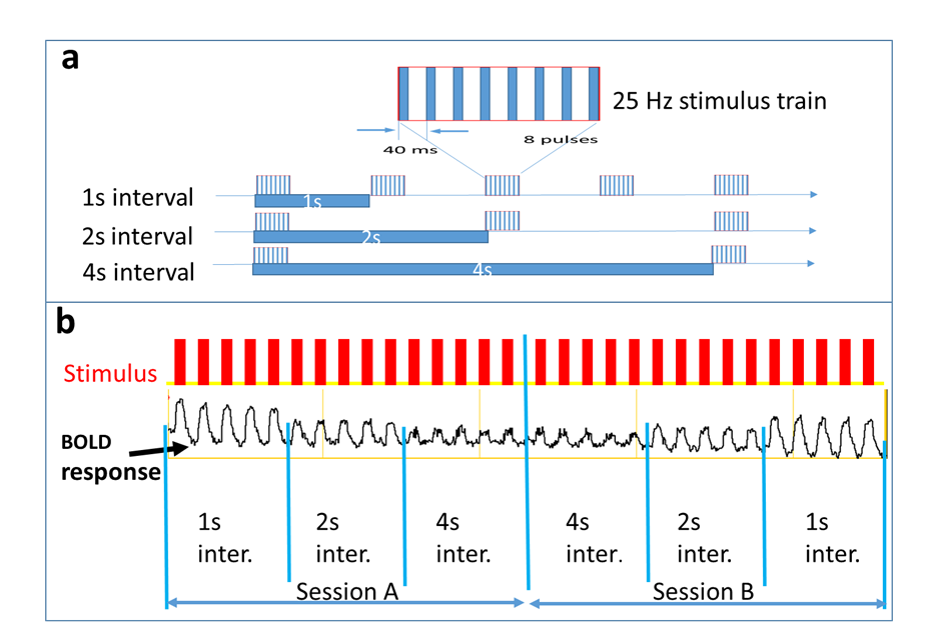
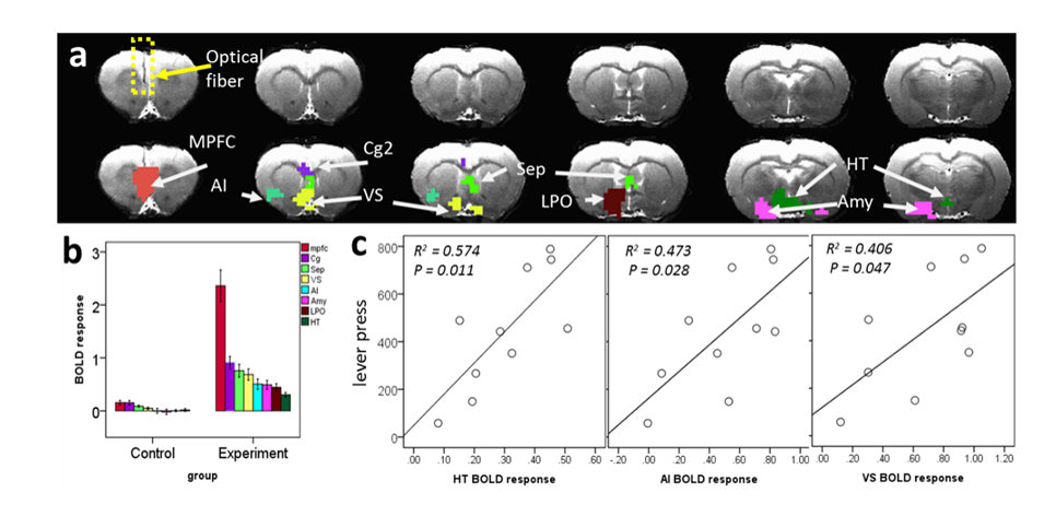
We injected channelrodopin2-expressing adeno-associated virus (AAV-ChR2) or control virus (AAV-eYFP) into the infralimbic region of the MPFC of experimental (n=10) or control (n=7) rats, respectively. Animals were trained to press a lever for optogenetic self-stimulation. A stimulation train consisted of 8 pulses (3ms duration) paced every 40 ms was delivered every time when the animal pressed the lever. Animals were subsequently anesthetized with a combination of low dose of isoflurane and dexmedetomidine,6 and underwent fMRI scanning on a Bruker 9.4T scanner. Block-design optogenetic stimulation was delivered to the right MPFC under three conditions: 25Hz stimulus trains with an interval of 1s, 2s, or 4s, respectively (Fig 1a). Each condition consisted of 5 blocks, and each block consisted of 20s stimulus on and 40s off. Two scan sessions with the stimulus order of A) 1s-2s-4s-interval or B) 4s-2s-1s-intervel were performed (Fig 1b). The order of scans A and B was counterbalanced between animals. BOLD fMRI data were acquired using a T2*-weighted EPI sequence (TE = 13 ms, TR = 1000 ms, segment = 2, FOV = 35 × 35 mm2, matrix size = 64×64, slice thickness = 1 mm, slice number = 15). Brain activation was analyzed with general linear modeling. Relationship between the fMRI activation and reward behavior was examined using a correlation analysis.Results
Lever press (within 30-minute sessions) assessed outside of scanner indicated that the experimental rats showed higher response to the stimulation than the controls (409±252 and 19±13, respectively; p= 0.00042, Fig 1b). The amplitude of BOLD response was modulated by the stimulus interval (higher amplitude at shorter interval) and was independent of the stimulus order (Fig 2b). Stimulation of MPFC activated many areas along known projections of the MPFC (p < 0.05, corrected for multiple comparisons; Fig 3). Specifically, the entire MPFC, including the medial orbital, prelimbic and infralimbic regions, was activated. The stimulation also activated cortical and subcortical regions that receive the MPFC afferents, including the agranular insula (AI), anterior cingulate area 2, tenia tecta, amygdala, ventral striatum (VS), septal area, bed nucleus of stria terminalis, midline anterior thalamic areas, preoptic area, and hypothalamus (HT). As shown in Fig 4, the lever press behavior was positively correlated with brain activation under the 1s-interval condition in the HT (r=0.76, p=0.01), AI (r=0.69, p=0.028) and VS (r=0.64, p=0.047).Discussion
Our study revealed that stimulation of the MPFC activates extensive brain regions, and some of the activation was correlated with the lever press behavior. The infralimbic region of rat MPFC corresponds to the human subcallosal cingulate,7 which is an effective target of deep brain stimulation for the treatment of depression.1, 2 Our finding may shed light on brain circuits involved in therapeutic effects of DBS.
Acknowledgements
This work was supported by the Intramural Research Program of the National Institute on Drug Abuse.References
1. H. S. Mayberg, Targeted electrode-based modulation of neural circuits for depression. J Clin Invest 119, 717-725 (2009).
2. C. Hamani et al., The subcallosal cingulate gyrus in the context of major depression. Biol Psychiatry 69, 301-308 (2011).
3. A. Routtenberg, M. Sloan, Self-stimulation in the frontal cortex of rattus norvegicus. Behavioral Biology 7, 567-572 (1972).
4. D. Corbett, A. Laferriere, P. M. Milner, Elimination of medial prefrontal cortex self-stimulation following transection of efferents to the sulcal cortex in the rat. Physiology and Behavior 29, 425-431 (1982).
5. Z. B. You, T. M. Tzschentke, E. Brodin, R. A. Wise, Electrical stimulation of the prefrontal cortex increases cholecystokinin, glutamate, and dopamine release in the nucleus accumbens: an in vivo microdialysis study in freely moving rats. J Neurosci 18, 6492-6500 (1998).
6. H. Lu et al., Rat brains also have a default mode network. Proceedings of the National Academy of Sciences of the United States of America 109, 3979-3984 (2012).
7. S. R. Heilbronner, J. Rodriguez-Romaguera, G. J. Quirk, H. J. Groenewegen, S. N. Haber, Circuit-Based Corticostriatal Homologies Between Rat and Primate. Biol Psychiatry 80, 509-521 (2016).
Figures