1151
3D MRI of Knee in Pediatric Patients with CAIPIRINHA SPACE: Diagnostic Performance Assessment with Arthroscopic Correlation1The Johns Hopkins University School of Medicine, Baltimore, MD, United States, 2Siemens Healthcare GmbH, 3Siemens Healthcare USA
Synopsis
3D CAIPIRINHA SPACE permits the acquisition of 4-fold accelerated, high quality data sets and has been shown to be feasible for efficient 3D MRI of the knee; however, the clinical application has not been demonstrated. We report the performance of 3D CAIPIRINHA SPACE MRI for the diagnosis of internal derangement of the knee in children and adolescents using arthroscopy correlation as the standard of reference. 3D CAIPIRINHA SPACE enables clinically feasible isotopic 3D MRI of the knee in children and adolescents with an acquisition time of 10 min and high accuracy for the diagnosis of meniscal, ligamentous and cartilage abnormalities.
Introduction
While the MRI evaluation of internal derangement of the knee relies heavily on two-dimensional (2D) turbo spin echo (TSE) pulse sequences, the separate acquisitions of MR images of different image planes is a time consuming process. Three-dimensional (3D) TSE sequences, such as Sampling Perfection with Application optimized Contrast using different flip angle Evolutions (SPACE), in contrast, can acquire isotopic data sets which high spatial resolution that afford reconstruction of any image planes from a single volume data set [1]. The combination of CAIPIRINHA (Controlled Aliasing In Parallel Imaging Results IN Higher Acceleration) and SPACE permits the acquisition of 4-fold accelerated, high quality data sets through undersampling in both the phase and partition encoding directions and optimized use of coil sensitivities, which mitigates aliasing artifacts with high acceleration factors [2]. 3D CAIPIRINHA SPACE has been shown to be feasible for 3D MRI of the knee with pulse sequence acquisition times of 5:00 min and less achieving image quality similar than conventional 3D SPACE and 2D TSE MRI [3]; however, the clinical application has not been reported.Purpose
We prospectively assessed the performance of 3D CAIPIRINHA SPACE MRI for the diagnosis of internal derangement of the knee in children and adolescents using arthroscopy correlation as the standard of reference.Methods
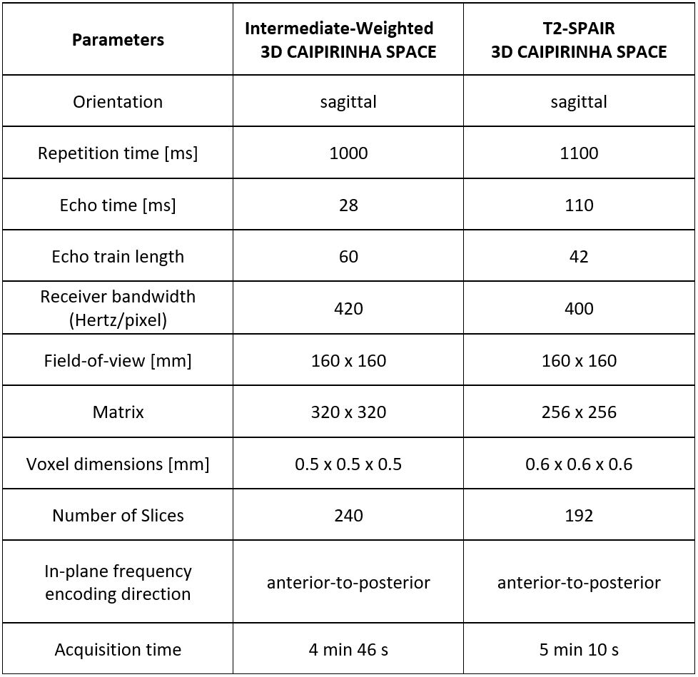
Institutional review board approval was obtained and informed consent was obtained. Twenty symptomatic subjects (12 boys, 8 girls; mean age, 12 years; age range, 6–16 years) underwent 3T MRI of the knee using a 3T MRI system (MAGNETOM Skyra, Siemens Healthcare, Erlangen, Germany) and a 1-channel transmit/15 channel receive coil (QED, Mayfield, OH). The MRI protocol consisted of isotropic intermediate-weighted and fat-suppressed T2SPAIR 3D CAIPIRINHA SPACE sequence prototypes (Figure 1) with a total acquisition time of 10 minutes. All patients underwent arthroscopic surgery within 38 (1-132) days after the MRI by a fellowship-trained orthopedic pediatric surgeon. Following anonymization and randomization, the 3D data sets were evaluated by two fellowship-trained, full time musculoskeletal radiologists in an independent fashion using diagnostic work stations and a predefined hanging protocol with interactive multiplanar reconstruction mode. The degree of motion and overall diagnostic quality was graded with equidistant Likert scales. Further, the presence or absence of discoid menisci was assessed, as well as the integrity of the menisci, cartilage, anterior cruciate ligament. Menisci were graded as intact or torn. Cartilage was graded as intact or defect. The anterior cruciate ligament was graded as intact or torn. During arthroscopy, the anatomic structures were systematically examined and graded using the same criteria. The data of the two observers were combined to increase statistical power. Mean sensitivity, specificity, positive and negative predictive value, and accuracy of 3D CAIPIRINHA SPACE was calculated.Results
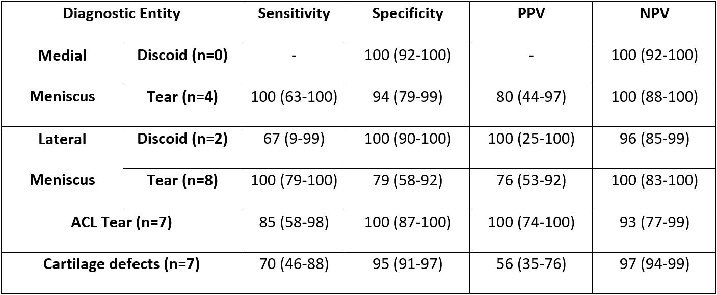
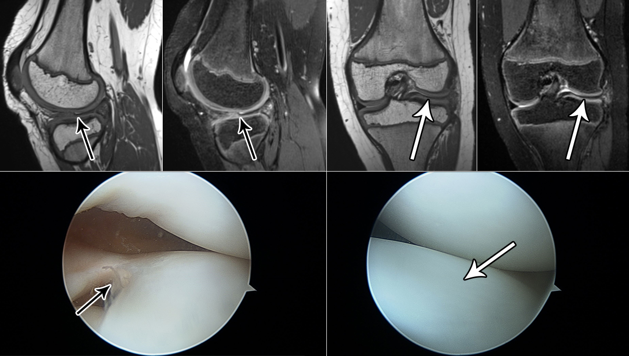
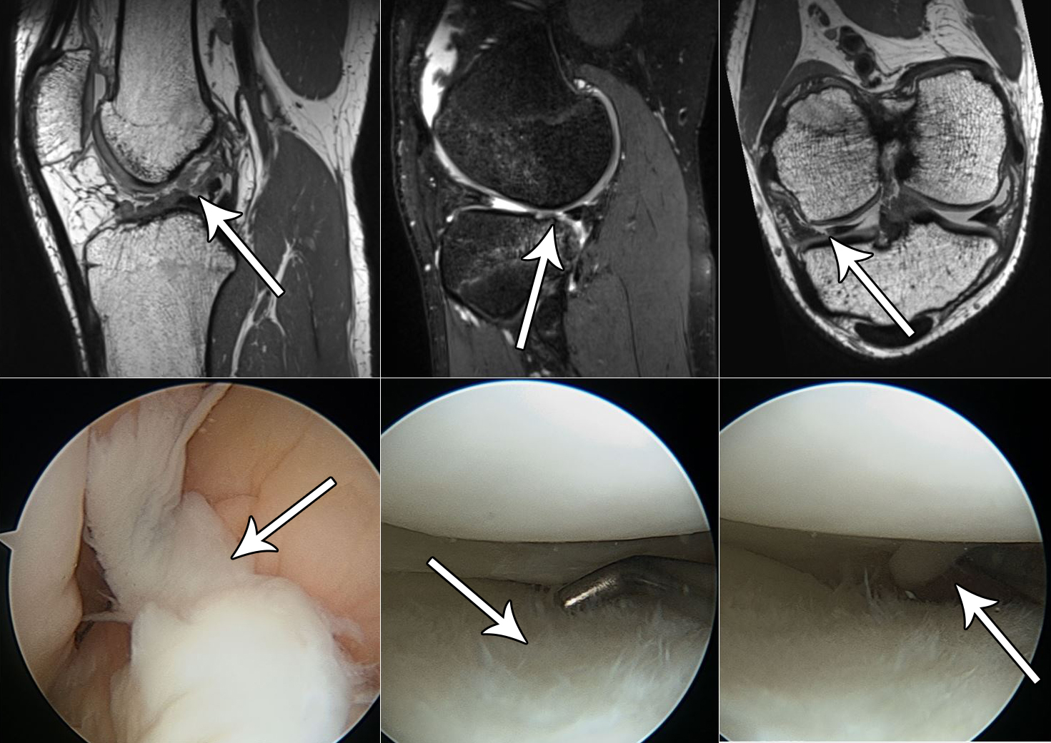
3D CAIPIRINHA SPACE MRI of the knee was successfully completed in all 20 subjects. Two studies demonstrated mild and moderate degradation of image quality by motion with a high interrater agreement kappa coefficient of 0.773 (95%-CI, 0.438 – 1.000). The overall diagnostic quality was rated very good (adequate – very good) with high interrater agreement kappa coefficient of 0.722 (0.388 – 1.000). There were 2 lateral discoid menisci and no medial discoid menisci (Figure 3). There were 4 medial and 8 lateral meniscal tears (Figure 4). Seven anterior cruciate ligament tears were diagnosed by arthroscopy and there were 7 cartilage defects (Figure 4). The diagnostic performance parameters are given in Figure 2.Discussion
Our data demonstrate the clinical applicability of 3D CAIPRINIHA SPACE for the assessment of internal derangement of the knee in children and adolescents with an acquisition time of 10 min. The advantages of the 2-dimensional 2x2 CAIPIRINHA pattern compared to a 1-dimensional acceleration pattern are the elimination of aliasing artifacts and of centrally located image noise [3]. Our initial results demonstrate sensitivities between 67-100% as well as specificities of 79-100% for the diagnosis of meniscal, intra-articular ligamentous and articular cartilage abnormalities. Further studies with larger sample sizes are ultimately needed to narrow the confidence intervals of the performance parameters.Conclusion
3D CAIPIRINHA SPACE enables clinically feasible isotopic 3D MRI of the knee in children and adolescents with an acquisition time of 10 min and high accuracy for the diagnosis of meniscal, ligamentous and cartilage abnormalities.Acknowledgements
We thank Qiong Zhang for his work on the sequence prototype.References
1. Mugler JP. Optimized three-dimensional fast-spin-echo MRI. J Magn Reson Imaging. 2014 Apr;39(4):745-67. doi: 10.1002/jmri.24542. Epub 2014 Jan 8.
2. Breuer FA, Blaimer M, Mueller MF, et al. Controlled aliasing in volumetric parallel imaging (2D CAIPIRINHA). Magn Reson Med 2006; 55:549-556.
3. Fritz J, Fritz B, Thawait GG, Meyer H, Gilson WD, Raithel E. Three-Dimensional CAIPIRINHA SPACE TSE for 5-Minute High-Resolution MRI of the Knee. Invest Radiol. 2016 Oct;51(10):609-17.
Figures