1150
Rapid knee MRI using TSE sequences accelerated with a combination of simultaneous multislice, multicoil compressed sensing and elongated echo trains1Center for Advanced Imaging Innovation and Research (CAI2R), NYU School Of Medicine, New York, NY, United States, 2Biomedical Engineering, School of Engineering, New York University, Brooklyn, NY, United States, 3Center for Biomedical Imaging, Dept of Radiology, NYU School Of Medicine, New York, NY, United States, 4Department of Radiology, NYU School Of Medicine, New York, NY, United States
Synopsis
Turbo spin echo imaging is accelerated with a novel combination of simultaneous multislice, multicoil compressed sensing and elongated echo times to reduce the scan time of routine knee examinations to 7 minutes. The accelerated protocol is validated against the conventional technique in a cohort of 10 patients based on the opinion of expert radiologists. Diagnostic accuracy was found to be similar despite the reduction in scan time. This might be useful to expand the utilization of MRI and to increase patient throughput.
Purpose
To develop accelerated 2D Turbo Spin Echo (TSE) sequences of the knee using a combination of simultaneous multislice (SMS) (1,2), multicoil compressed sensing (SPARSE-SENSE) (3,4) and elongated echo trains in order to decrease exam time of routine knee MR examinations while preserving image contrast, SNR and resolution for diagnostic accuracy.
Materials and Methods
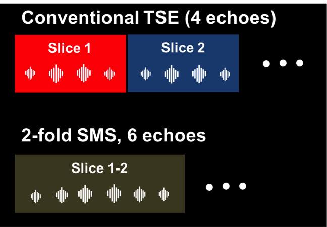
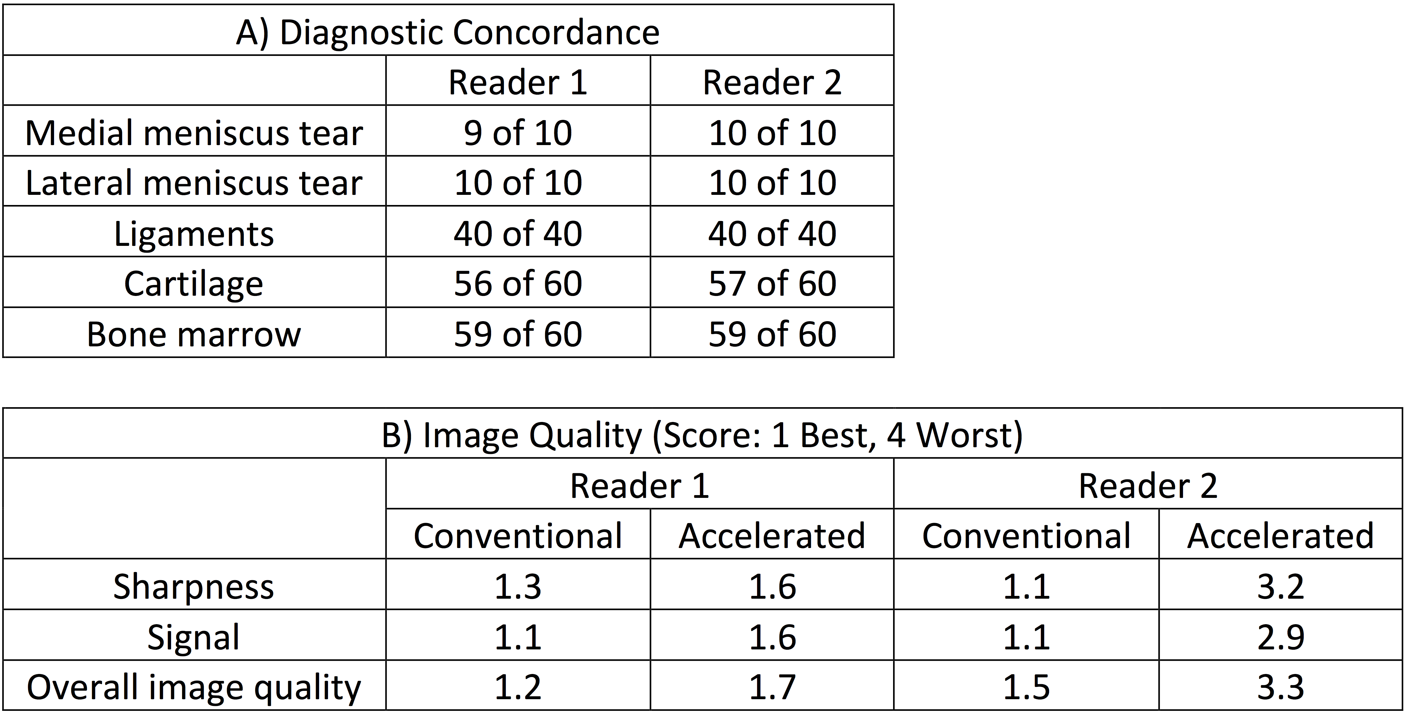
A multislice TSE pulse sequence was modified to introduce SMS acceleration along the slice dimension and SPARSE-SENSE (compressed sensing and parallel imaging) acceleration along the phase-encoding dimension. The latter was implemented using variable-density random undersampling based on a Poisson disk distribution and following the same echo train (ET) division pattern in k-space as the clinical sequence to preserve contrast. Initial attempts to utilize SMS to accelerate the sequence by decreasing TR proved unsuccessful due to loss of optimal contrast, blurring and decreased SNR. Therefore, it was decided to leave TR unchanged and take advantage of the acceleration of SMS by lengthening the ET (Fig.1). Following optimization of the sequence, 10 subjects were enrolled in the study (approved by the IRB). Each subject underwent imaging with our conventional protocol for internal derangement of the knee as well as the proposed accelerated protocol. The protocols consisted of sagittal PD and FS T2, coronal PD and FS PD, axial FS T2 2D TSE sequences. The conventional used GRAPPA acceleration R=2 (TA=11.42min) and the accelerated protocol used SMS=2, SPARSE-SENSE= 2, and elongated ET (TA = 7.12min). The ET was increased by 50% for PD sequences and 18-22% for T2 sequences from the conventional to the accelerated protocol. Joint SMS and SPARSE-SENSE reconstruction was performed offline in Matlab by introducing the SMS model in the data acquisition operator and enforcing multicoil image compressibility in the wavelet domain. The MRIs of the 10 subjects were reviewed by two fellowship trained musculoskeletal radiologists (experience from 1-16 years) blinded to MRI acquisition and reconstruction parameters. Cases were reviewed in two different sessions, to minimize recall bias. MRIs were assessed for meniscal tears, ligament tears, bone marrow signal abnormalities, and articular cartilage defects. Criteria were binary indicators of whether or not each reader detected specific pathologies and subjective (reader dependent) 4-point ordinal assessment of overall image quality.
Results
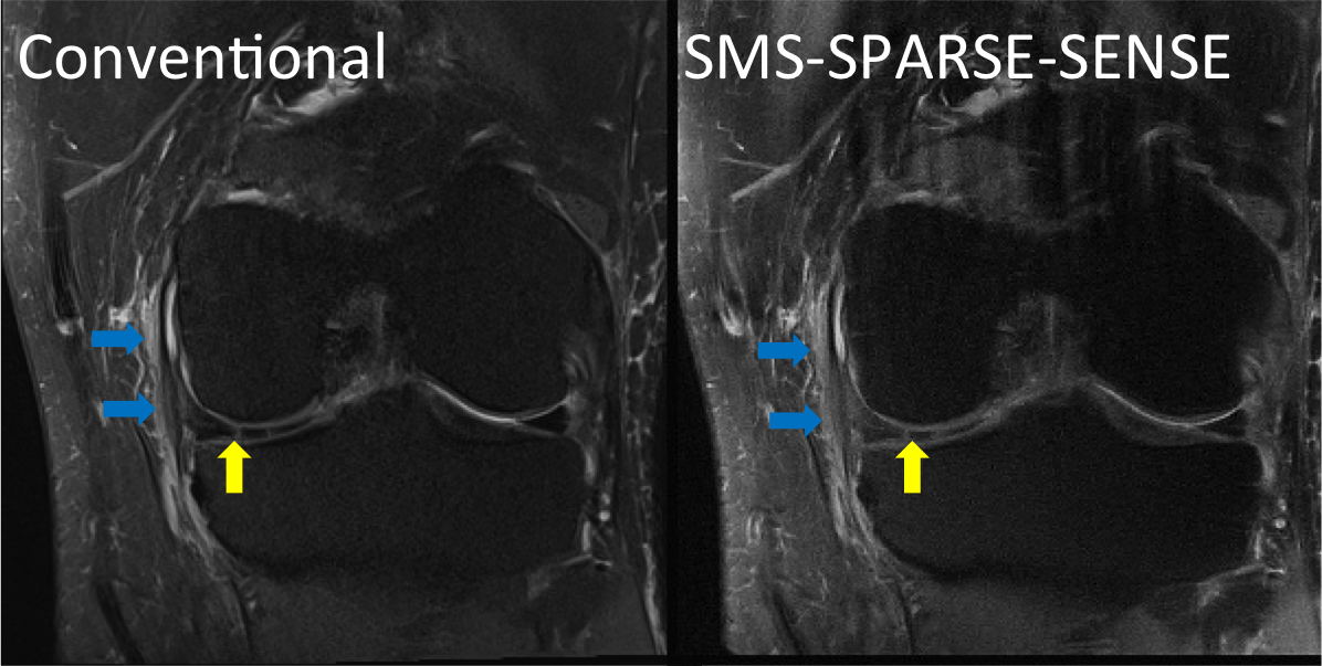
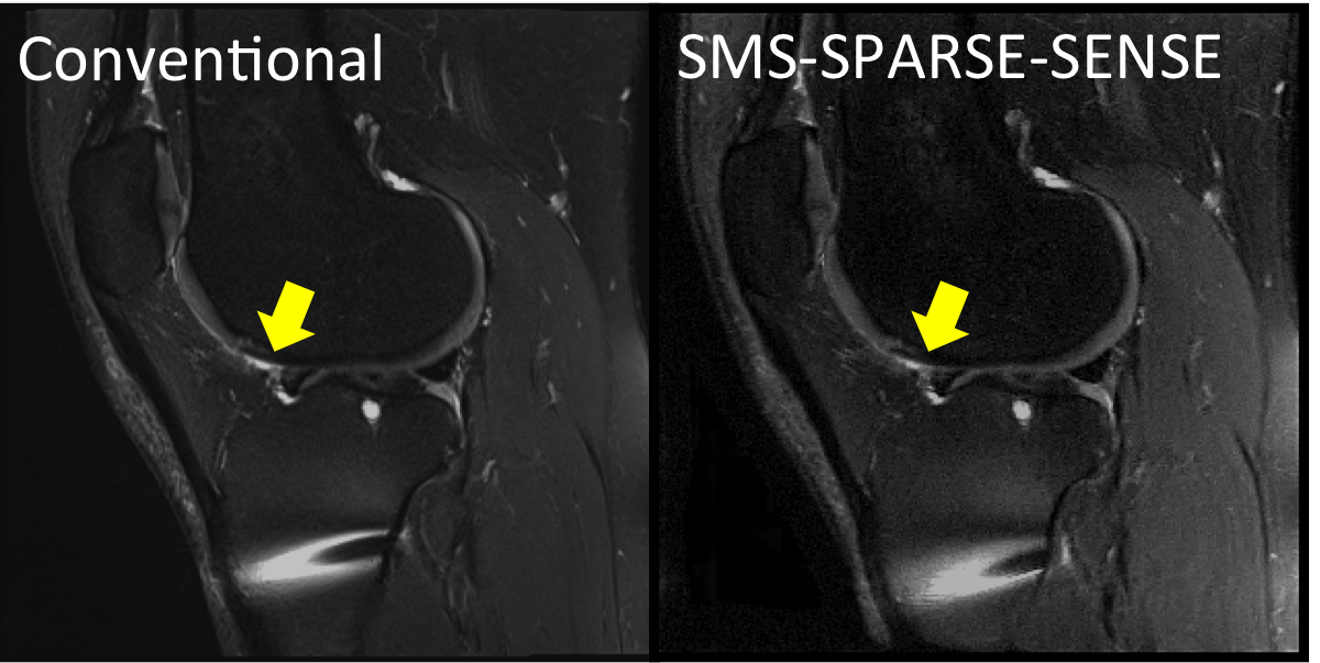
SMS-SPARSE-SENSE with 4-fold data reduction and elongated ET decreased total examination time by a factor of 1.6 with respect to the conventional protocol (2-fold parallel imaging acceleration). Diagnostic accuracy of both techniques was found to be similar by both readers, while image quality received mixed reviews (Figure 2). Reader 1 found a small decrease in image quality while reader 2 reported about 2-fold decrease in image quality. This perceived image quality decrease is possibly due in part to degradations in SNR caused by shorter acquisition times and resolution due to the SPARSE-SENSE reconstruction. Specific cases showing different lesions are shown in Figures 3-5, where the proposed SMS-SPARSE-SENSE method depicts similar clinical information content with respect to the clinical protocol.Discussion
A new application of SMS is demonstrated for rapid TSE imaging, where acceleration is achieved without changes in contrast by increasing echo train length instead of the common approach of reducing the TR. This approach effectively reduces the number of TRs. The combination of SMS along the slice dimension and in-plane SPARSE-SENSE optimizes the use of the CAIPIRINHA shifts (2) and improves performance over in-plane SENSE or GRAPPA. If 2-fold SENSE or GRAPPA acceleration was used in plane, only a CAIPIRINHA shift of ¼ FOV would be possible, since the FOV was already reduced by a factor 2. The random undersampling pattern in SPARSE-SENSE evenly distributes the undersampling artifacts over the whole FOV and thus enables to still use a ½ FOV shift and improve the overall performance of SMS. The decrease in signal to noise and resolution observed by one of the readers is likely due to longer ET and the relatively lower compressibility of 2D images. Future work will try to address this issue by including a T2 decay model in the reconstruction, which may also enable longer ET.
Conclusion
The synergetic combination of SMS, SPARSE-SENSE and elongated ET enables a 7-minute routine knee examination with similar diagnostic accuracy to the conventional 11.4-minute protocol. This might be particularly useful to expand the utilization of MRI and to increase patient throughput.
Acknowledgements
Funding: 1P41EB017183-01A1References
[1] Barth, Markus, et al. "Simultaneous multislice (SMS) imaging techniques."Magnetic resonance in medicine 75.1 (2016): 63-81.
[2] Breuer, Felix A., et al. "Controlled aliasing in parallel imaging results in higher acceleration (CAIPIRINHA) for multi-slice imaging." Magnetic resonance in medicine 53.3 (2005): 684-691.
[3] Lustig, Michael, David Donoho, and John M. Pauly. "Sparse MRI: The application of compressed sensing for rapid MR imaging." Magnetic resonance in medicine 58.6 (2007): 1182-1195.
[4] Otazo, Ricardo, et al. "Combination of compressed sensing and parallel imaging for highly accelerated first-pass cardiac perfusion MRI." Magnetic Resonance in Medicine 64.3 (2010): 767-776.
Figures