1129
Lighter is better: A Flexible Lightweight Eight Channel Slot Antenna Array for Cardiac MRI at 7.0 Tesla1Berlin Ultrahigh Field Facility (B.U.F.F.), Max Delbrück Center for Molecular Medicine in the Helmholtz Association, Berlin, Germany, 2MRI.TOOLS GmbH, Berlin, Germany, 3Experimental and Clinical Research Center (ECRC), a joint cooperation between the Charité Medical Faculty and the Max Delbrück Center for Molecular Medicine in the Helmholtz Assosciation, Berlin, Germany, 4DZHK (German Centre for Cardiovascular Research, partner site Berlin, Germany
Synopsis
The wave length in tissue at ultrahigh fields allows for practical realization of RF antenna architectures such as dipole elements, high dielectric resonators and slot antennae. This work presents a novel flexible eight channel slot antenna array customized for cardiac MRI at 7.0 T. The proposed array is lightweight, easy to build, and affords a tight fit for a broad range of upper torso geometries while ensuring good matching and tuning. The in vivo study demonstrated the feasibility of the array for high fidelity, whole heart coverage MRI at 7.0 T and showed rather uniform signal intensity across the heart.
Introduction
The wave length (λ) shortening in tissue at
ultrahigh fields (B0≥7.0T) permits antenna architectures like dipole
antennae [1, 2] and
slot antennae [3] to
be used in transmit receive arrays in practical sizes. Recent studies
demonstrated that compared to a dipole antenna of the same size, a single slot
antenna can provide a higher SNR with lower RF power deposition. Notwithstanding
this success, the extension of a λ/2 slot antenna (λ/2 = 50 cm at 300 MHz)
constitutes a severe challenge for realization of two-dimensional arrays covering
the upper torso. In light of this constraint, we propose in this work a
flexible eight channel slot antenna array for cardiac imaging at 7.0 T, which
is lightweight, easy to build and cost effective. The array was designed using
numerical field simulations, and the RF performance of the array was evaluated
in phantom studies. The feasibility of this slotted array for high resolution imaging
with whole heart coverage was demonstrated in a pilot in vivo study in healthy subjects.Methods
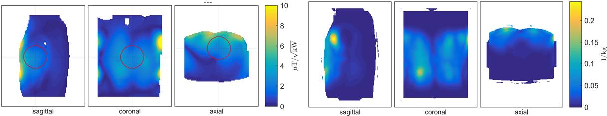
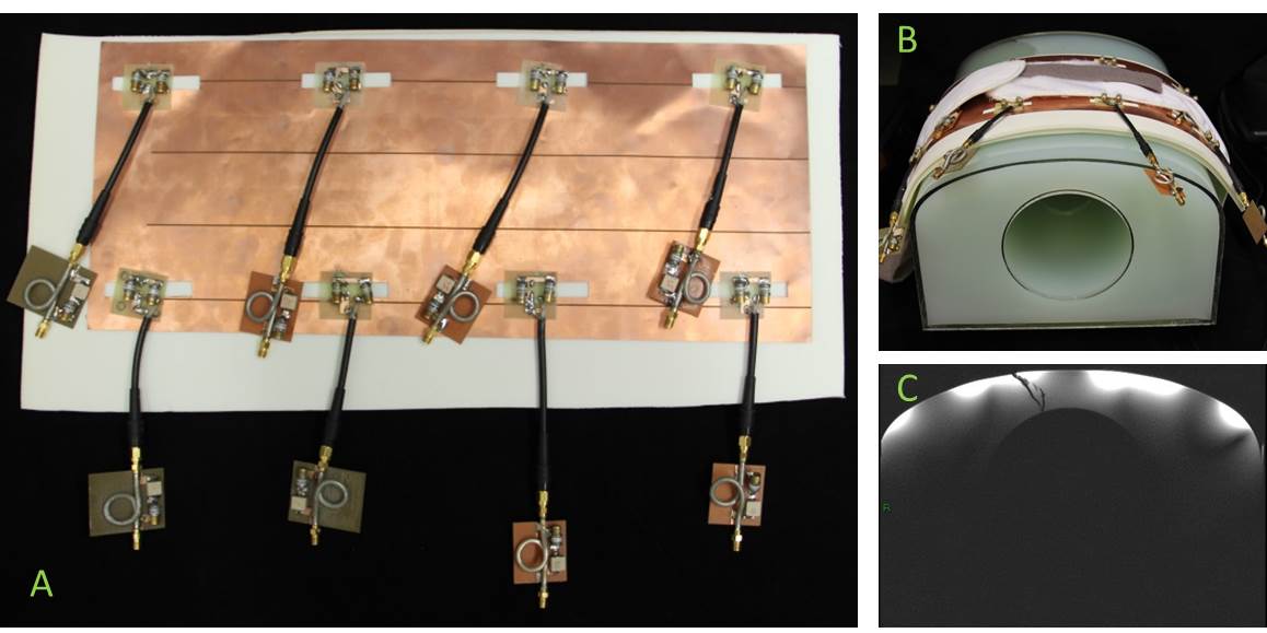
Electromagnetic field simulations were performed using CST Studio Suite 2015 (CST AG, Darmstadt, Germany) with a human voxel model Duke (BMI: 23.1 kg/m2) from the Virtual Family to assess the geometry, safety and B1+ performance of the slot antenna array (Fig.1 left). Based on this, we designed an array with 8 elements in a 2x4 configu
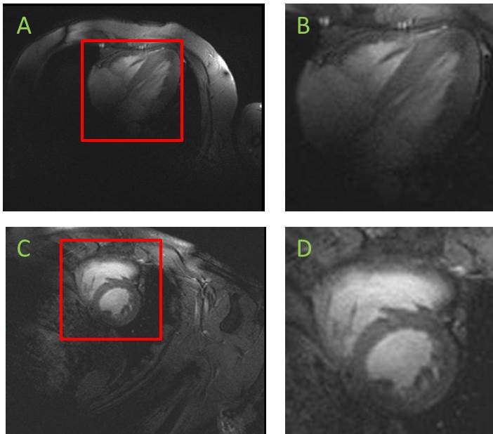
ration (Fig.1 right). With this arrangement the channels of the array are decoupled only by distance, achieving a balance between the number of channels and the size of the array. The array was constructed on a single flexible sheet of copper by cutting out the slot holes. A matching and tuning circuit was placed at the feeding point of each slot (Fig 3.). To reduce the SAR, a 20 mm flexible padding was incorporated beneath the array. The array was matched and tuned on a torso phantom filled with dielectric gel (ε=75 σ=0.73 S/m). S-parameter matrix was acquired on the same phantom using an 8 channel network analyser (ZVT 8, Rohde & Schwarz, Memmingen, Germany). In vivo imaging experiments were performed using a 7.0 T whole body MR system (Magnetom, Siemens, Erlangen, Germany). Four chamber (4CV) and short axis view (SAX) images of the heart were acquired using single breath hold 2D CINE FLASH (matrix = 290 x 352, in-plane spatial resolution = (1.0 x 1.0) mm2, slice thickness = 4mm, TR = 6.29 ms, TE = 2.96 ms). A tight yet comfortable fit of the array for the volunteer was provided by wrapping the array around the torso. For a homogeneous excitation no additional phase delay (phase setting) between the channels was needed.
Results
The proposed flexible slot antenna array is lightweight (m=475 g) including the feeding cables sitting on the anterior torso, the cable traps and the tuning and matching circuits. Due to its flexibility the array conforms to a broad range of upper torso geometries. With a height of 30 mm, the proposed array consumes minimal space in the effective magnet bore, which enables cardiac MR of subjects covering an ample BMI range. The slot antenna array yielded reflection coefficients better than -18 dB. The highest coupling between the elements was -11 dB. Reflection coefficients and coupling did not change substantially when the bending of the array was changed. SAR values, derived from the EM simulations incorporating a male human voxel model were well below the limits permitted by the IEC guidelines [4] for an average accepted input power of 10 W. 2D CINE FLASH imaging of the heart provided high spatial resolution images (1.0 x 1.0 x 4.0) mm2 with good blood-myocardium contrast (Fig. 4). Given the blood-myocardium contrast, the anatomic coverage and the transmission field uniformity of this proposed array, small structures such as valve cups and trabeculae carneae can be depicted.Conclusion
Our preliminary results demonstrate the feasibility of an 8 channel slot antenna array for cardiac imaging at 7.0 T, which provides high resolution images with whole heart coverage, without major signal voids in the target region. The lightweight, small height and flexibility of the slot antenna array architecture offers superior patient comfort, while maintaining good matching, tuning and image quality. Due to the easy construction the slot antenna array design presents an appealing RF antenna alternative for further applications or regions of interest such as abdomen, knee, head and spine by modifying the number, size and arrangement of the slots of the array.Acknowledgements
No acknowledgement found.References
1. Oezerdem, C., et al., 16-channel bow tie antenna transceiver array for cardiac MR at 7.0 tesla. Magnetic Resonance in Medicine, 2016. 75(6): p. 2553-2565.
2. Raaijmakers, A., et al., Design of a radiative surface coil array element at 7 T: The single side adapted dipole antenna. Magn Reson Med, 2011. 66(5): p. 1488-1497.
3. Leeor Alon, C.M.D., Ryan Brown, Daniel Sodickson, Christopher M. Collins. A Slot Antenna Concept for High Fidelity Body Imaging at Ultra High Field. in ISMRM 2016. 2016. Singapore.
4. Commission, I.E., Medical electrical equipment. Part 2-33: Particular requirements for the safety of magnetic resonance equipment for medical diagnosis, in IEC 60601-2-33, IEC: Geneva.
Figures