1126
An 8/15-Channel Tx/Rx Head Neck RF Coil Combination with Semi-Dynamic B1 Shimming for Improved fMRI of the Cerebellum at 7 T1Erwin L. Hahn Institute for Magnetic Resonance Imaging, University Duisburg-Essen, Essen, Germany, 2High Field and Hybrid MR Imaging, University Hospital Essen, Essen, Germany, 3Department of Neurology, University Hospital Essen, Essen, Germany
Synopsis
Functional MRI of the human cerebellum is challenging at ultrahigh fields, since conventional RF head coils hardly cover the cerebellum with sufficient signal-to-noise ratio and B1+-inhomogeneities introduce challenges. In order to overcome these problems, a coil combination consisting of an 8ch transceiver head coil and a 7ch receive only array are combined to improve imaging of the whole brain with special focus on the cerebellum. A ‘semi-dynamic’ B1+-shimming technique is introduced which provides a tSNR-gain of 29 % and voxels with higher significance in a finger tapping fMRI experiment when comparing the coil combination to a 32ch receive head coil.
Purpose:
Functional
magnetic resonance imaging (fMRI) at 7 T benefits from higher signal-to-noise
ratio (SNR) as well as increased functional contrast based on blood oxygenation
level. To fully exploit these advantages, radiofrequency (RF) coils need to
provide sufficient signal at the region of interest (ROI). Furthermore, B1+-inhomogeneities
need to be addressed to ensure a homogeneous excitation. In this work, the main
ROI is the human cerebellum. Its high anatomical diversity and location pose
challenges for fMRI1. In order to overcome these, we combine an 8ch
transceiver (Tx/Rx) head coil2 with a 7ch receive only array3 including
modified detuning to provide high local SNR at the cerebellum. ‘Semi-dynamic’ B1+-shimming
was employed in combination with Variable Slice-Thickness (VAST) EPI4,
to perform fMRI focusing on the human cerebellum, while maintaining whole brain
coverage. The RF-shimming method’s performance was evaluated in the coil combination
setup and compared with a conventional 1chTx/32chRx coil (Nova Medical Inc.,
Wilmington, USA) in terms of B1+-homogeneity and temporal
signal-to-noise ratio (tSNR) during a basic finger tapping fMRI experiment.Methods:
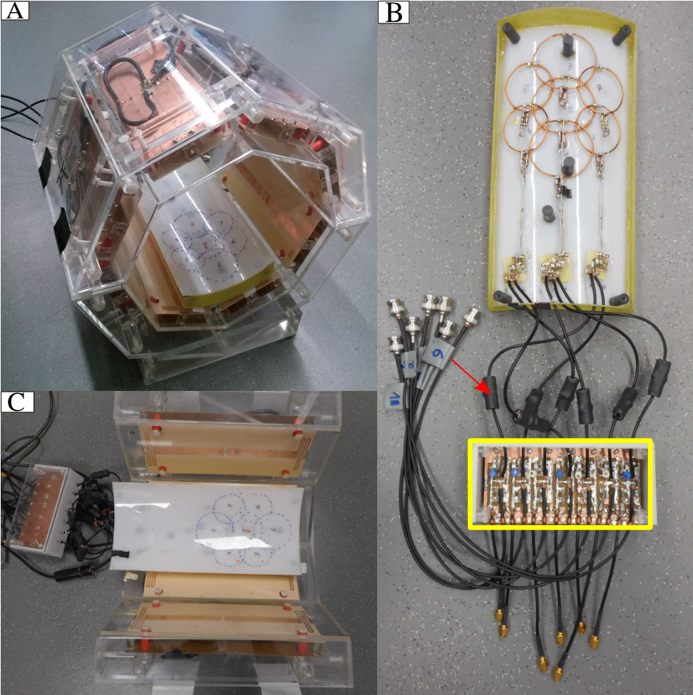
The coil combination setup as depicted in fig. 1A & C consists of the 8ch Tx/Rx coil in which the 7ch receive array is placed occipital. A detuning network (fig. 1B yellow box) consisting of seven circuits with PIN diodes was implemented to improve detuning. The coil was additionally equipped with floating cable traps (fig. 1B red arrow) to decrease coupling between the receive array and the transceiver coil. In an in vivo acquisition (healthy male volunteer: 35 y, 75 kg, 175 cm) with the coil combination, flip angle (FA) maps for four slices with a thickness of 24 mm (fig. 2A) were obtained using B1TIAMO5. Based on this information, three different RF-shims were calculated (fig. 2A). The VAST-EPI sequence was modified by introducing an additional trigger which control the switching between the individual shim settings via a custom modulator system6. In fig. 3A, the red and blue slice groups represent an acquisition with 1.5 mm slice thickness, where the red block is shimmed with one setting (fig. 2B) and the blue block with another (fig. 2C). The yellow slice block is acquired with 3 mm slice thickness and shimmed with the third setting (fig. 2D). The same participant was scanned with the Nova coil, whereas dielectric bags where used to passively shim the B1+-field for the cerebellum. With these shimming procedures, finger tapping experiments with a random tapping pattern of the right hand (TE/TR = 22/2800 ms, TA = 9:41 min, 28 s tapping vs. 28 s rest in ten blocks resulting in 202 volumes) were conducted. Functional t-maps in the cerebellum and neocortex where obtained using SPM12 and the SUIT7 toolbox for cerebellum only. As a measure of quality, tSNR-maps in the cerebellum were calculated using the first hundred realigned functional scans. Therefore, the voxel-wise mean and standard deviation along the time course where calculated and divided by each other.Results and Discussion:
In fig. 2B-D, the resulting FA maps after RF-shimming are shown. From left to right, they correspond to the three slice positions from bottom to top shown in fig. 2A, whereas fig. 2E-G shows maps of the Nova birdcage mode. This result indicates a FA increase and improved homogeneity, especially at the cerebellar position. Since the experiment is driven by a hypothesis of the location of activation, the shimming ROIs (black ROIs in fig. 2) are chosen a priori to homogenize B1+ for this particular area. This shimming process translates into higher significance when performing fMRI experiments (fig. 3B-D). However, significant caudal activation is less for the semi-dynamic shimming case in this particular experiment. One possible reason could be that the Nova coil fits the shape of the participant’s head better. The tSNR-maps for the cerebellum as shown in fig. 4 indicate higher tSNR with the coil combination (B) and a further increase when applying semi-dynamic shimming (C). Computing the mean tSNR in every slice and through all slices from caudal (most left) to cranial (most right) reveals a tSNR-gain of 16 % when applying semi-dynamic shimming in comparison to birdcage-mode of the coil combination and a gain of 29 % in comparison with the Nova coil.Conclusion:
A coil combination and method has been presented which improves fMRI of the human cerebellum in comparison with a conventional RF head coil, while maintaining whole brain coverage. An overall tSNR-gain in the cerebellum can be seen, although some parts seem to be covered less good with the receive elements. In future work, this experiment will be repeated in more participants applying group analysis.Acknowledgements
No acknowledgement found.References
1. J. Diedrichsen et al.: Advances in functional imaging of the human cerebellum. Curr. Opin. Neurol. 23(4). 382-7 (2010).
2. S. Orzada et al.: 8-channel transmit/receive head coil for 7 T human imaging using intrinsically decoupled strip line elements with meanders. Proc. 17th Annual Meeting ISMRM. #3010 (2009).
3. S. Rietsch et al.: Cost-Efficient 7ch Rx Shoulder Array for 7T UHF MRI Featuring External Switchbox Detuning. Proc. 24th Annual Meeting ISMRM. #2142 (2016).
4. S. Brunheim et al.: Variable Slice-Thickness (VAST) EPI for the reduction of signal voids in GE-EPI at 7 Tesla Proc. Org. Hum. Brain Map. 20. #2089 (2014).
5. S. Brunheim et al.: Combining B1 mapping with TIAMO for fast and accurate multi-channel RF shimming in 7 Tesla body MRI. Proc. 24th Annual Meeting ISMRM. #936 (2016).
6. A.K. Bitz, I. Brote, S. Orzada et al.: An 8-channel add-on RF shimming system for whole-body 7 Tesla MRI including real-time SAR monitoring. Proc. 17th Sci. Meet. Int. Soc. Magn. Reson. Med. #4767 (2009).
7. J. Diedrichsen et al.: A spatially unbiased atlas template of the human cerebellum. Neuroimage 33(1). 127-138 (2006).
Figures