1080
Functional and structural connectivity of the cingulate bundle related to future cognitive performance in MS1Imaging Institute, The Cleveland Clinic, Cleveland, OH, United States, 2HemaImaging, 3Neurological Institute, The Cleveland Clinic, Cleveland, OH, United States, 4Center for Brain Health, The Cleveland Clinic, Cleveland, OH, United States
Synopsis
This work assesses the relationship of resting state fMRI (rs-fMRI) and DTI of the posterior cingulum bundle to future cognitive performance. We find that rs-fMRI is related to performance on measures related to episodic memory, and radial diffusivity is related to performance on a measure of speed of processing.
Introduction
Over 40% of Multiple Sclerosis (MS) patients suffer from cognitive dysfunction [1], though due to the diffuse nature of the disease the neural basis of cognitive decline is unclear. Our previous work found a relationship between white matter integrity and information processing speed and visual spatial episodic recall in the posterior portion of the cingulum bundle (PCB) [2]. As part of a longitudinal study assessing the sensitivity of pathway-specific imaging measures in MS, we present preliminary data on the relationship of longitudinal resting state functional connectivity (rs-fMRI) and radial diffusivity (RD) in the cingulum bundle to cognitive performance after two years.Methods
In an IRB-approved protocol, seven patients with MS (age: 51.71±7.6, 1 male, EDSS: 3.86±1.6) were scanned at 6 time-points over the course of two years. All participants completed neuropsychological testing at visit 1 and visit 6, using counterbalanced equivalent forms for each measure. Tests included the SDMT, a measure of information processing speed, the CVLT-II, a measure of verbal episodic memory, and the BVMT-R, a measure of spatial episodic memory.
MRI acquisition: Scans were acquired at 3T using a 12-channel receive-only head array. Parameters were as follows: rs-fMRI: 132 repetitions of 31-4mm thick axial slices acquired with TE/TR=29ms/2800 ms, 128x128 matrix, 256mm x 256mm FOV, receive bandwidth=1954Hz/pixel, eyes closed. DTI: HARDI data were acquired with a twice-refocused spin echo [3] (TE/TR=102/7700msec, 128x128x48 matrix, FOV=256x256x96mm), 71 b=1000 sec/mm2 acquisitions with gradient directions selected by a coulomb repulsion algorithm [4], and 8 b=0 acquisitions at equally spaced intervals.
Data analysis: Rs-fMRI scans were corrected for motion and physiologic noise, detrended, and lowpass filtered [5,6]. Using a previously described method [7], nine-voxel in-plane seeds were placed bilaterally in the posterior cingulate cortex and hippocampus. With each hemisphere, the timeseries from the two seeds were correlated. The normalized correlation [8] was averaged across hemispheres and taken as a measure of the strength of rs-fMRI in the PCB.
DTI analysis is described in reference [2]. The seed regions defined above were used as seed (posterior cingulate cortex) and target (hippocampus) regions for probabilistic fiber tracking, as described in [2]. Pathway-dependent RD was calculated using the track density map, the scalar diffusion values, and a white matter mask and averaged across left and right hemispheres.
Statistical analysis: RD and rs-fMRI of the PCB were correlated with cognitive measures at Times 1 and 6 and the percent change in cognition for each subject. Imaging and cognitive measures at Times 1 and 6 were compared with paired Student’s t-tests.
Results
Rs-fMRI in the PCB was lower at Time 6 as compared to Time 1 (p = 0.005). RD was higher, though not significantly so (p = 0.072). Performance on the SDMT decreased significantly from Time 1 to Time 6 (p = 0.012). CVLT and BVMT performance also decreased, though these tests did not reach significance.
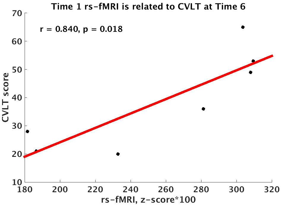
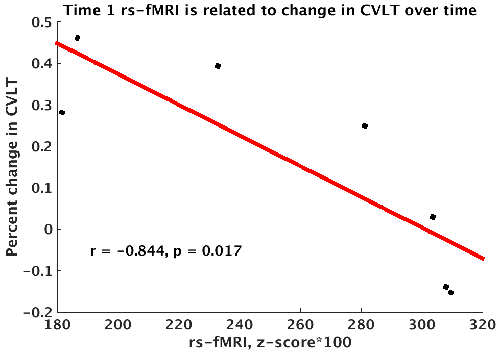
Rs-fMRI at Time 1 was weakly related to performance on the BVMT at Time 1 (r = 0.685, p = 0.090), and was more strongly related to Time 2 performance on the BVMT (r = 0.902, p = 0.006), and CVLT (r = 0.840, p = 0.018; Figure 1). Rs-fMRI at Time 1 was also related to the change in performance over time on the CVLT (r = -0.844, p = 0.017; Figure 2) and was weakly related to change on the BVMT (r = -0.716, p = 0.070).
RD at Time 1 was related to performance on the SDMT at both Time 1 (r = -0.786, p = 0.036) and Time 6 (r = -0.873, p = 0.010). RD at Time 1 was not related to the change in SDMT performance over time.
Discussion and Conclusion
With a limited sample, we find that the relationship of both structural and functional connectivity is related to cognitive performance over time in MS. In particular, rs-fMRI measures seem to be more strongly related to later cognitive performance. Increased PCB connectivity at Time 1 is related to both better performance on episodic memory measures at Time 6 and to a smaller decline in cognitive measures. The stronger relationship of rs-fMRI to future cognitive performance may relate to disparate rs-fMRI findings in the literature. Data from additional subjects are currently being collected and will allow more sophisticated statistical analysis of the predictive ability of rs-fMRI with regard to cognitive decline.Acknowledgements
This work was supported by National Multiple Sclerosis Society grant number RG4931A1.References
1. Achiron A, Chapman J, Magalashvili D, Dolev M, Lavie M, Bercovich E, et al. Modeling of Cognitive Impairment by Disease Duration in Multiple Sclerosis: A Cross-Sectional Study. PLoS One. 2013;8(8):e71058
2. Koenig, K.A., Sakaie, K.E., Lowe, M.J., Lin, J., Stone, L., Bermel, R.A., Beall, E.B., Rao, S.M., Trapp, B.D., Phillips, M.D. The relationship between cognitive function and high-resolution diffusion tensor MRI of the cingulum bundle in multiple sclerosis. Multiple Sclerosis Journal. 2015 Jun 23. pii: 1352458515576983. [Epub ahead of print] PubMed PMID: 26106010
3. Reese, TG, Heid, O, Weisskoff, RM, Wedeen, VJ: Reduction of eddy-current-induced distortion in diffusion MRI using a twice-refocused spin echo. Magn Reson Med. 2003; 49:177-82, (2003).
4. Jones, DK, Horsfield, MA, Simmons, A: Optimal strategies for measuring diffusion in anisotropic systems by magnetic resonance imaging. Magn Reson Med. 1999; 42:515-25.
5. Beall EB and Lowe MJ. SimPACE: generating simulated motion corrupted BOLD data with synthetic-navigated acquisition for the development and evaluation of SLOMOCO: a new, highly effective slicewise motion correction. Neuroimage. 2014; 101:21-34.
6. Glover G, Li T, Ress D. Image-Based Method for Retrospective Correction of Physiological Motion Effects in fMRI: RETROICOR. Magnetic Resonance in Medicine 2000; 44:162-67.
7. Lowe, MJ, Koenig, KA, Beall, EB, Sakaie, KA, Stone, L, Bermel, R, Phillips, MD: Anatomic connectivity assessed using pathway radial diffusivity is related to functional connectivity in monosynaptic pathways. Brain connectivity. 2014; 4: 558-65.
8. Lowe MJ, Mock BJ, Sorenson JA. Functional connectivity in single and multislice echoplanar imaging using resting-state fluctuations. NeuroImage. 1998; 7(2):119-32.
Figures