1078
Altered effective connectivity of dorsolateral prefrontal cortex in obsessive-compulsive disorder: a Granger causality analysis with resting-state fMRI1Huaxi Magnetic Resonance Research Centre(HMRRC),West China Hospital of Sichuan University, Chengdu, People's Republic of China
Synopsis
In current study, we demonstrated the altered effective connectivity between bilateral dorsolateral prefrontal cortex(DLPFC) and other brain regions in obsessive compulsive disorder(OCD) using the Granger causality analysis of resting-state functional MRI. We found that the effective connectivity from the bilateral DLPFC to some brain regions are increased or decreased in OCD ,and DLPFC is the important information flow center. We also observed positive correlations between the abnormal causal effect and clinical symptoms such as obsession. These findings provide insight into OCD-related neural network disorders and may potentially guide clinical diagnosis and treatment of OCD in the future.
Purpose
Obsessive-compulsive disorder(OCD) is a common psychiatric characterized by the presence of obsessions and compulsions. Previous study has suggested its cerebral pathological model of the dysfunction in dorsolateral prefronto-striatal loop[1]. It is assumed that the deficit in DLPFC-related functional connectivity was associated with the cause of the impairment of spatial attention and working memory processes in OCD [2]. However, as functional connectivity is a synchronous, non-directed correlation, the direction of information flow and causal effect between DLPFC and other regions of the brain remains unclear. Here, we aimed to investigate the directionality of information transfer and causal effect from the DLPFC to the other brain regions, as well as the feedback effect from the other regions to the DLPFC. We further investigated whether those effective connectivities would correlate to the severity of clinical symptoms as measured by scores such as Yale-Brown Obsessive Compulsive Scores(Y-BOCS) and Hamilton Anxiety Scale(HAMA).Materials and Methods
The study was approved by the Ethical committee of West China Hospital of Sichuan University, and written informed consent was obtained from each participant. A total of 88 OCD patients and 88 sex and age matched HC participated in the present study(Table 1). OCD patients were recruited from the Mental Health Center of West China Hospital and diagnoses were confirmed by using the Structured Clinical Interview for DSM-IV.
All MR data were collected with a 3-Telsa GE MRI imaging system. Resting-state functional MR were obtained using a GE-EPI sequence with below parameters: TR/TE=2000/30msec,FOV 240×240mm,flip angle=90°,slice thickness=5mm with no gap,30 axial slices,200 volumes in each run. The functional MRI data were pre-processed using SPM8(http://www.fil.ion.ucl.ac.uk/spm) and Data Processing Assistant for Resting-state fMRI(DPARSFA, http://rfmri.org/DPARSF). We performed bivariate voxel-wise Granger Causality Analysis(GCA) using the REST-GCA in the REST toolbox (http://www.restfmri.net).
We chose bilateral DLPFC as two individual seeds based on the coordinates by Matthew JH et al[3](bilateral DLPFC: x=±56, y=26, z=25, Radius=4mm).OCD and HC were compared by two-sample t test with SPM and a FDR-corrected p value of less than 0.05 at cluster-level was deemed to be significant. To examine the correlations between the abnormal causal effect and clinical variables, Pearson correlation were performed between these variables.
Results
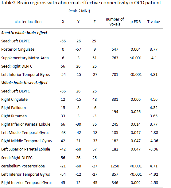
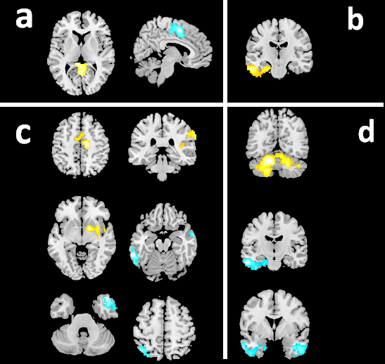
From DLPFC to the whole brain: Compared with HC,OCD patients showed increased or excitatory effective connectivity from the left DLPFC to the posterior cingulate and from the right DLPFC to the left inferior temporal gyrus(ITG). While decreased or inhibitory effective connectivity were found from the left DLPFC to the supplementary motor area(SMA)(Table 2 and Figure 1).
From whole brain to DLPFC: Compared with HC, OCD patients showed increased or excitatory effective connectivity from the right cingulate, right pallidum, right putamen and right inferior parietal lobule(IPL)to the left DLPFC. While the decreased or inhibitory effective connectivity were found from bilateral middle temporal gyrus(MTG) and left superior parietal lobule(SPL) to the left DLPFC in OCD patients. In addition, the excitatory effective connectivity from posterior lobe of cerebellum to the right DLPFC and the inhibitory effective connectivity from the bilateral ITG to the right DLPFC were also found in OCD patients.
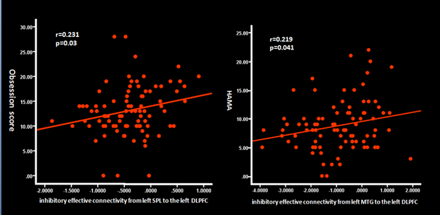
Correlations between between abnormal effective connectivity and clinical variables: As shown in Figure 2, positive correlation was found between the inhibitory effective connectivity from the left MTG to the left DLPFC and the HAMA score in OCD patients(r=0.21,p=0.041). The inhibitory effective connectivity from the left SPL to the left DLPFC was positively associated with obsession scores in Y-BOCS(r=0.23,p=0.03).
Discussion&Conclusion
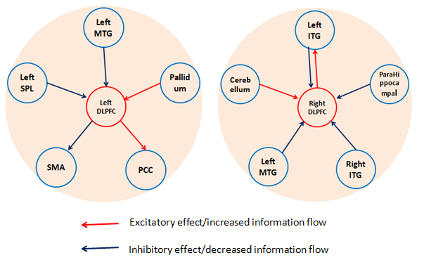
The main findings of our study is the altered excitatory/ inhibitory effective connectivity between the DLPFC and other brain regions, which could also be defined as the increased/decreased information inflow or outflow between the cortical regions. Our study may demonstrate that DLPFC is the important information flow center(Figure 3). DLPFC could excite or inhibit the function of some regions(such as PCC,SMA and left ITG),and it can also be influenced by other regions(such as pallidum, putamen, MTG, IPL,ITG and cerebellum).We also observed positive correlations between abnormal effective connectivity and clinical symptom severities which prove its disease related role. These findings are important for studying the highly directional neurological system in psychiatric disorders such as OCD and can help us understand the underline mechanism from a neural network prospect. We hope that these findings may in-depth our understanding of the disorder and provide more target treatment of OCD in the future. Further study is needed to investigate whether those altered effective connectivity is associated with structural deficits.Acknowledgements
This study was supported by the National Natural Science Foundation (Grant No. 81671669 and 81171488),and Chinese college students innovation training program.References
[1]Menzies,L et al. Neurosci Biobehav Rev. 2008;32(3):525-49.
[2]TogaoO, et al. Psychiatry Res. 2010 Oct 30;184(1):29-37.
[3] Hoptman MJ, et al. Schizophr Bull. 2010 Sep;36(5):1020-8
Figures
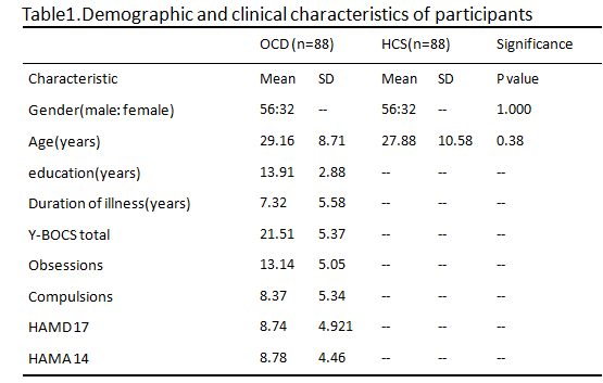
Table1.Demographic and clinical characteristics of participants.Abbreviations: HAMA, Hamilton Anxiety Rating Scale; HAMD, Hamilton Depression Rating Scale; HCS, healthy control subjects; OCD, obsessive–compulsive disorder; SD, standard deviation; Y-BOCS, Yale-Brown Obsessive Compulsive Scale