1077
Dissociation of structural and functional dysconnectivity in first-episode drug-naive schizophrenia1Radiology, Huaxi MR Research Center, West China Hospital of Sichuan University, Chengdu, People's Republic of China
Synopsis
Combining DTI and fMRI data sets, the present study revealed the dissociation of structural and functional dysconnectivity in a large cohort of first-episode drug-naive schizophrenia patients.
Introduction
Functional and structural neuroimaging studies in schizophrenia both provide evidence of network dysconnectivity in widespread brain regions.1,2 However, the relationships between structural and functional connectivity alterations remain ill-defined in this disorder. According to previous reviews 1,3, the structural and functional alterations of brain regions including medial prefrontal cortex, superior temporal cortex, thalamus, striatum, anterior cingulate, and hippocampus were most frequently reported in first-episode drug-naive schizophrenia, where the effects of heterogeneous illness duration and treatment would not confound. This study focused analytical resources to these pre-described regions of interest to increase the chance of detecting potentially subtle alterations of structural and functional connectivity and their relationship in the early course schizophrenia by combining diffusion tensor imaging (DTI) and resting-state functional magnetic resonance imaging (fMRI) in a large cohort of first-episode drug-naive schizophrenia patients.Methods
DTI and fMRI data sets were acquired on a 3T MR system for 203 first-episode drug-naive schizophrenia patients and 185 healthy controls. Several diffusion parameters including fractional anisotropy, number, and length of fiber tracts derived from tensor-guided tractography were to quantify specific structural connectivity and temporal correlation coefficients using the Pearson correlation were to quantify functional connectivity in subjects’ native anatomical space. Group comparisons were performed for each modality to characterize structural and functional dysconnectivity in patients group and correlation analyses were performed to test structural-functional relationships by correlating diffusion parameters with corresponding temporal synchrony coefficients of same regions, and with clinical symptom ratings.Results
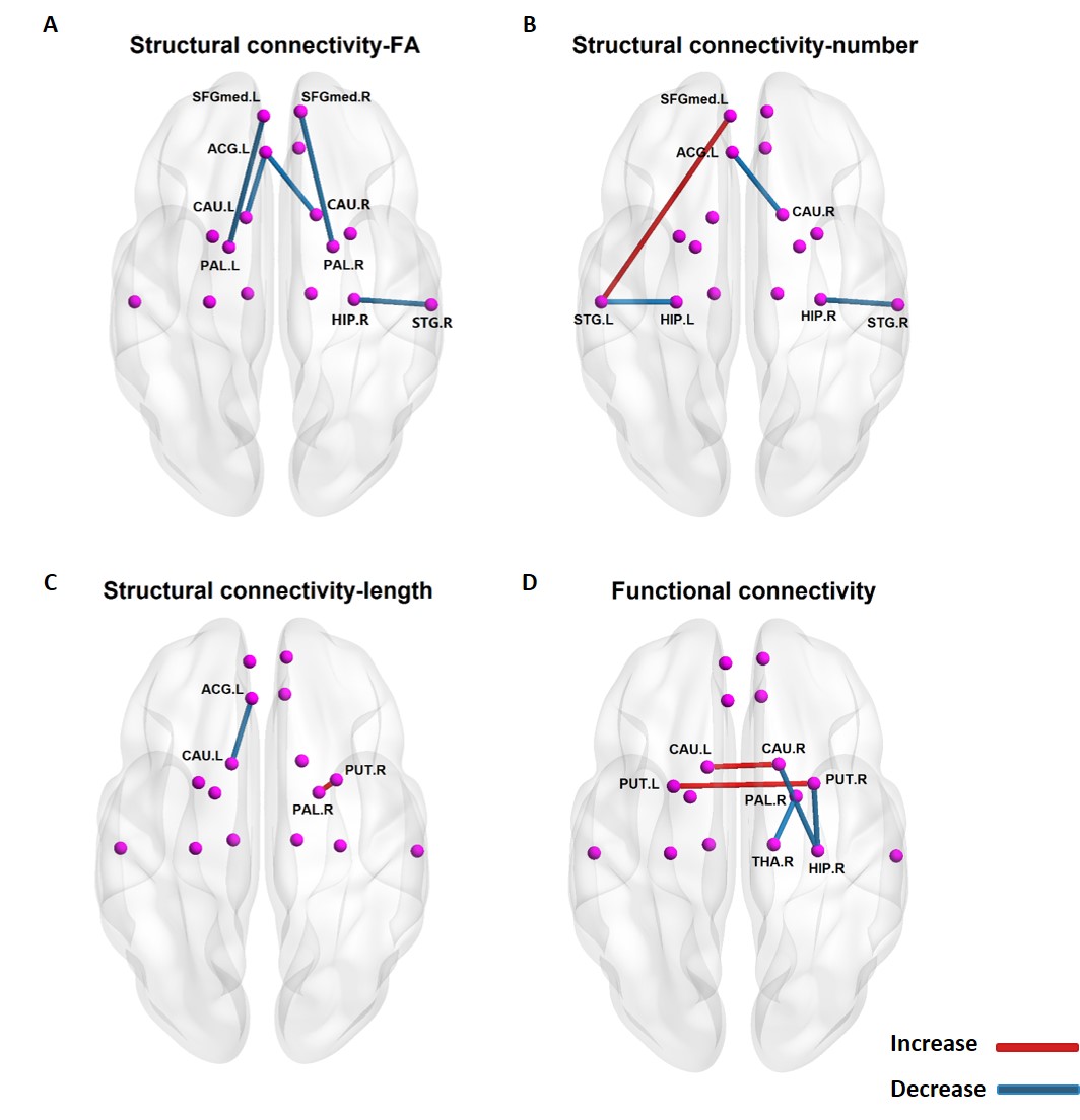
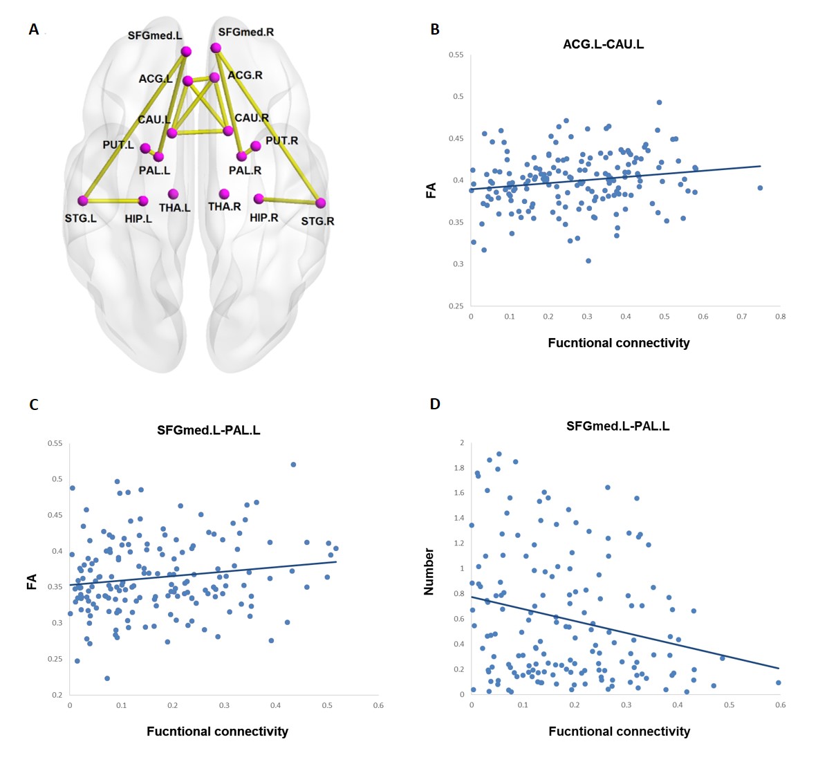
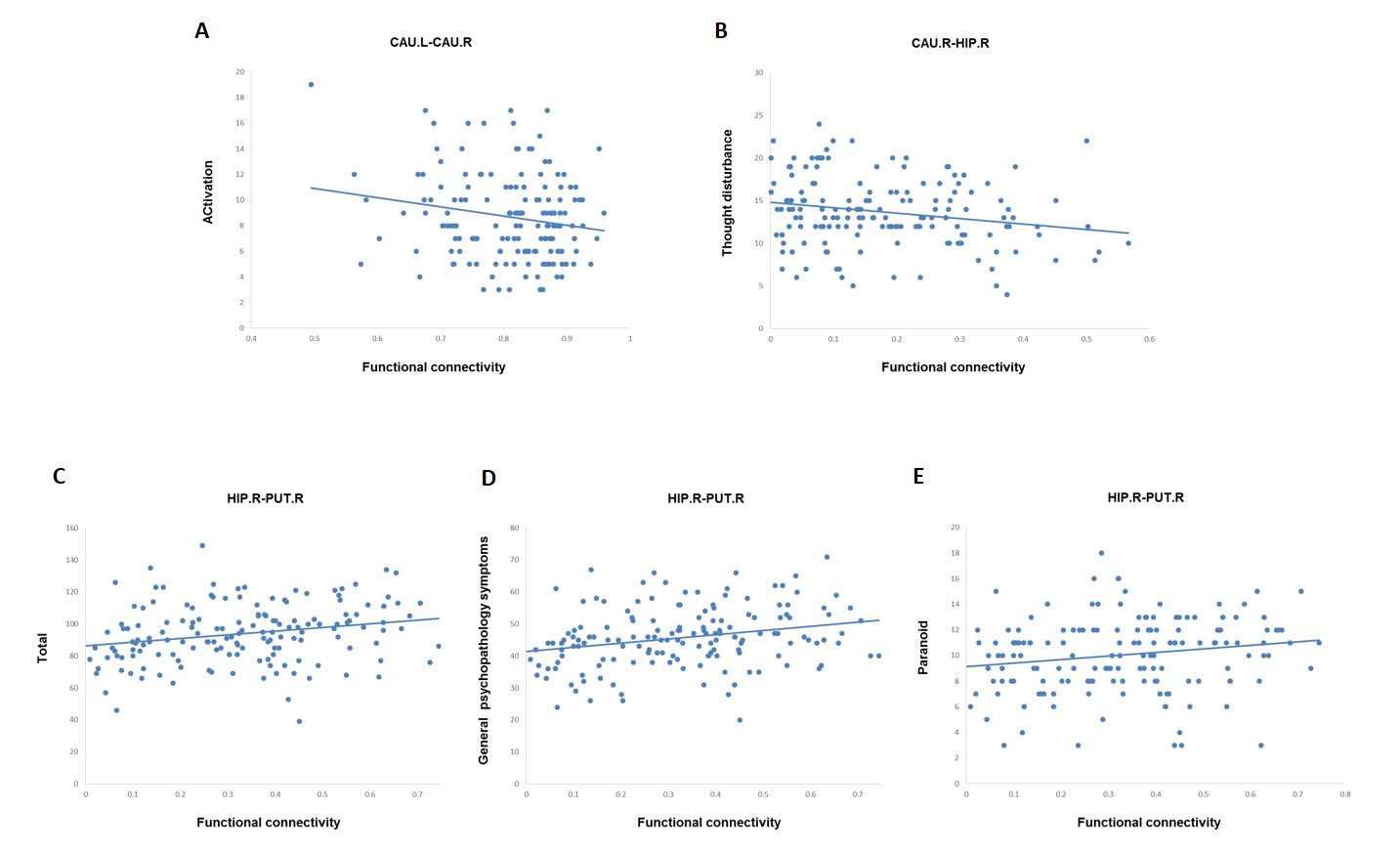
Group comparisons revealed both structural and functional dysconnectivity in the patients. Structural dysconnectivity were mainly found between medial prefrontal cortex and pallidum, anterior cingulate gyrus and caudate, and superior temporal gyrus and hippocampus while functional dysconnectivity were found between bilateral caudate, bilateral putamen, right caudate and right hippocampus, right pallidum and right thalamus and right putamen and right hippocampus. Correlation analyses only found moderate structural-functional correlation between left anterior cingulate and left caudate nucleus as well as left medial prefrontal cortex and left pallidum in the patients. Besides, only functional dysconnectivity was correlated with symptom severity, though these associations were modest.Discussion
The most important finding was that group comparisons analysis revealed both structural and functional dysconnectivity in the patients. However, there was no regional overlap in these connections, suggesting the dissociation of structural and functional dysconnectivity within these brain regions. While multimodal studies in schizophrenia suggested a structural basis for abnormal functional connectivity 4,5, however, regions that are not directly connected by white matter tracts can be functionally connected.6 For example, individuals with developmental agenesis of the corpus callosum demonstrate robust inter-hemispheric functional connectivity.7 Besides, multiple lines of evidence support a neural basis for functional connectivity measured at rest.8 Previous electrical cortical recording study found that the patterns of brain activity elicited by cortical stimulation correspond to functional connectivity.9 Pharmacological study also found that modulation of the glutamatergic system via blockage of N-methyl-d-aspartate receptor altered functional connectivity.10 These evidence indicated that while white matter connectivity provided neural substrate for functional connectivity, functional dysconnectivity was not constrained to structural connection. The localized and moderate structural-functional correlation further illustrated a much more complicate relationship rather than linear correlation between the structural and functional dysconnectivity. It was also interesting to note that only functional dysconnecitvity showed significant correlation with the symptom severity, indicating the functional dysconnectivity more directly reflect psychosis-related neurophysiological changes.Conclusion
This study provides evidence that structural and functional dysconnectivity are largely dissociated in the early course of schizophrenia. While white matter connectivity provides neural substrate for functional connectivity, functional dysconnectivity is not constrained to structural connection and directly reflect psychosis-related symptoms at the early onset of schizophrenia.Acknowledgements
This work was supported by National Natural Science Foundation of China (grant numbers 81371527, 81671664, 81621003), National Program for Special Support of Eminent Professionals and National Program for Support of Top-notch Young Investigator.References
1. Pettersson-Yeo W, Allen P, Benetti S, et al. Dysconnectivity in schizophrenia: where are we now? Neurosci Biobehav Rev. 2011;35(5):1110-1124.
2. Sheffield JM, Barch DM. Cognition and resting-state functional connectivity in schizophrenia. Neurosci Biobehav Rev. 2016;61:108-120.
3. Gong Q, Lui S, Sweeney JA. A Selective Review of Cerebral Abnormalities in Patients With First-Episode Schizophrenia Before and After Treatment. Am J Psychiatry. 2016;173(3):232-243.
4. Marenco S, Stein JL, Savostyanova AA, et al. Investigation of anatomical thalamo-cortical connectivity and FMRI activation in schizophrenia. Neuropsychopharmacology. 2012;7(2):499-507.
5. Leroux E, Delcroix N, Dollfus S. Left fronto-temporal dysconnectivity within the language network in schizophrenia: an fMRI and DTI study. Psychiatry Res. 2014;223(3):261-267.
6. Honey CJ, Thivierge JP, Sporns O. Can structure predict function in the human brain? Neuroimage. 2010;52(3):766-776.
7. Owen JP, Li YO, Yang FG, et al. Resting-state networks and the functional connectome of the human brain in agenesis of the corpus callosum. Brain Connect. 2013;3(6):547-562.
8. Karbasforoushan H, Woodward ND. Resting-state networks in schizophrenia. Curr Top Med Chem. 2012;12(21):2404-2414.
9. He BJ, Snyder AZ, Zempel JM, et al. Electrophysiological correlates of the brain's intrinsic large-scale functional architecture. Proc Natl Acad Sci U S A. 2008;105(41):16039-16044.
10. Hoflich A, Hahn A, Kublbock M, et al. Ketamine-Induced Modulation of the Thalamo-Cortical Network in Healthy Volunteers As a Model for Schizophrenia. Int J Neuropsychopharmacol. 2015;18(9):1-11.
Figures