1043
Looping StarFlorian Wiesinger1, Anne Menini1, and Ana Beatriz Solana1
1GE Global Research, Munich, Germany
Synopsis
This abstract describes a novel imaging method called Looping Star. The unique imaging characteristics are 1) continuous 3D radial imaging with close to 100% sampling efficiency, 2) inaudible/silent scanning, and produces 3) multi-gradient echo images at equidistant echo times including an FID image at TE=0. Looping Star is demonstrated in phantom, and in-vivo experiments for T2* weighted imaging and T2* BOLD fMRI.
Purpose:
This abstract describes a novel imaging method called Looping Star [1-2]. The unique imaging characteristics are 1) continuous 3D radial imaging with close to 100% sampling efficiency, 2) inaudible/silent scanning, and produces 3) multi-gradient echo images at equidistant echo times including an FID image at TE=0. Looping Star is demonstrated in phantom, and in-vivo experiments for T2* weighted imaging and T2* BOLD fMRI.Methods:
The Looping Star pulse sequence (Fig. 1) is based on RUFIS-type [3] Zero TE imaging, with the gradient amplitude (red) kept constant and only directional gradient updates applied in between repetitions, resulting in silent 3D radial imaging. RF excitation (black) is achieved by very short block pulses; such that the RF excitation bandwidth covers the full imaging bandwidth. More specifically, from a top-down perspective, the pulse sequence is organized in Nseg number of segments, and each segment is divided into Nloop number of echo loops (Nloop=3 in Fig. 1A). Bottom-up, each loop consists of Nsp/loop self-refocused, 3D radial spokes (Nsp/loop=8 in Fig. 1A). RF excitation is active only for the first FID loop and turned-off afterwards. In this way, the later loops (magenta: 1st echo, green: 2nd echo) form gradient-echoes of the initial FID (blue) excited in the first loop. For each subsequent segment, the original spokes are rotated such as to provide uniform 3D k-space coverage (Fig. 1C). Because of minimal gradient switching and very short RF pulses, a sampling efficiency of close to 100% can be achieved. The Looping Star pulse sequence was implemented on a GE 3T MR750w scanner using a GEM head coil (both GE Healthcare, Waukesha, WI) and applied for T2* weighted and BOLD fMRI imaging. To minimize acoustic noise, gradient waveforms were smoothed, resulting in slightly bended 3D radial spokes. Image reconstruction was performed using an iterative, conjugate-gradient SENSE reconstruction, including TGV regularization and echo-in/out separation.Results:
Figure 2 illustrates in-vivo Looping Star (TE=32ms*[0, 1, 2, 3], Nloop=4, FA=1deg, BW=31.25kHz, FOV=192mm, 2mm isotropic resolution, Nsp/loop=16, scan time=80sec) with the four echo images depicted on the left and corresponding T2* and B0 off-resonance maps on the right. Signal from the skin and the mouth region rapidly disappears for the later echoes, owing to shorter T2* relaxation times but also destructive off-resonance blurring. Quantitative T2* measurements were performed in a uniform silicon oil phantom providing T2*~26ms consistent with a matched 3D gradient echo measurement. Figure 3 demonstrates Looping Star fMRI (TE=26.88ms*[0,1], Nloop=2, FA=1deg, BW=62.5kHz, FOV=192mm, 3mm isotropic resolution, Nsp/loop=32, scan time/volume=2.2s) for a simple finger-tapping task, providing 5-6% T2* BOLD signal change in the expected primary motor cortex. The obtained TE=26.88ms (top) and TE=0 (middle) echo images are clean of distortions. Accordingly, the BOLD activation maps can be directly overlaid on top of the anatomical T1-weighted scan (top) without separate distortion correction. For all the performed Looping Star experiments, the measured acoustic noise level was max 2dBA above ambient background noise.Discussion:
Zero TE provides native proton density information. Using inversion-recovery, or T2 preparation additional T1 and/or T2 contrast weighting can be added. The presented Looping Star method adds a gradient refocusing mechanism, thereby enabling T2* and B0 mapping as well as T2* BOLD fMRI. The silent imaging characteristic is important to improve patient comfort and/or reduce anxiety.Acknowledgements
No acknowledgement found.References
[1] Wiesinger et al, U.S. Patent 15/142,957 [2] Solana Sanchez et al, ISMRM 2016: p.104 [3] Madio et al, MRM 34(4): 525-529 (1995) [4] Solana Sanchez et al, ISMRM 2017: submitted.Figures
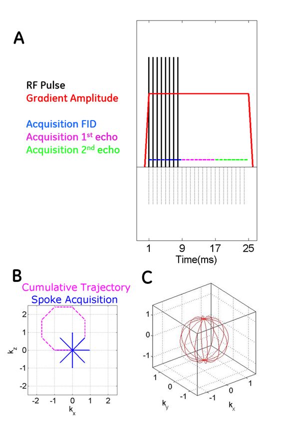
Figure 1: Looping Star
pulse sequence (top) and spokes arrangement (bottom left) resulting in gradient
refocusing of the original FID signal.
Via rotation full 3D k-space coverage is obtained (bottom right).
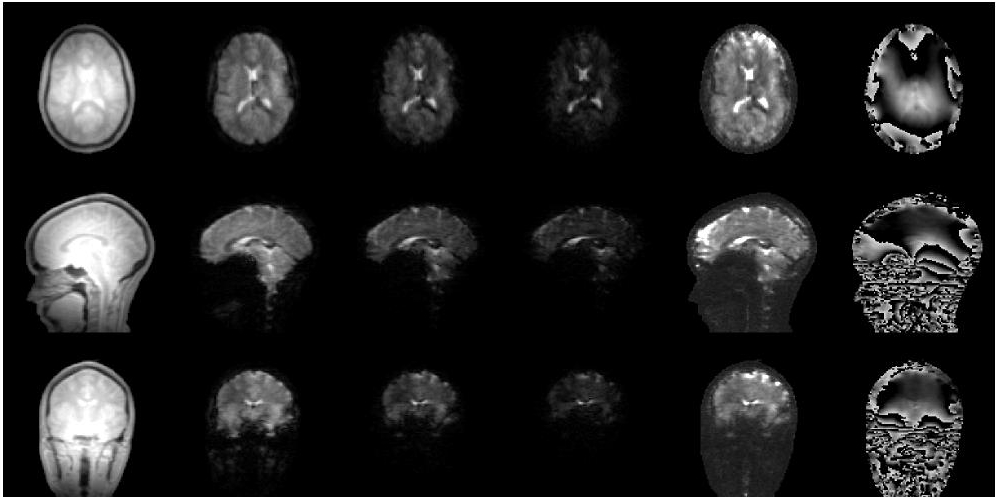
Figure 2: Looping Star TE=32ms*[0,
1, 2, 3] echo images (left) and corresponding T2* and B0 off-resonance map
(right).
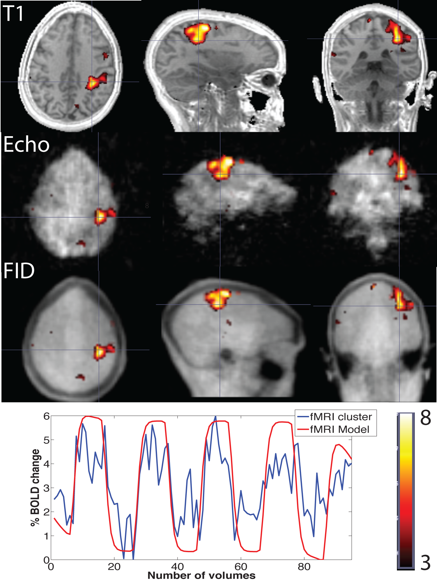
Figure 3: Looping
Star T2* BOLD fMRI using a finger tapping task. Because of negligible distortions, the BOLD
activation map can be directly overlaid on top of an anatomical T1w scans (top). The individual TE=26.88ms*[0,1] echo images
are shown in the middle. 5-6% T2* BOLD
signal change is obtained in the expected primary motor cortex.