1042
Single-Shot Spiral Arterial Spin Labeling MRI Enabled by Concurrent Field Monitoring1Institute of Biomedical Engineering, ETH Zurich, Zurich, Switzerland
Synopsis
Spiral k-space sampling holds great potential for many MRI applications particularly for arterial spin labeling which has inherently very low SNR. Besides providing significant increase in SNR by reducing the echo time and readout durations, spiral trajectories with single shot acquisitions are highly robust against motion artifacts. However their sensitivity to encoding deficiencies such as B0 off-resonance, field drifts, eddy currents, gradient coupling, gradient delays and concomitant fields prevents their utilization effectively in practice. In this work, we provide ASL with single-shot spiral readouts that are robust against all those deficiencies by using dynamic field monitoring concurrent with image acquisition.
INTRODUCTION
Perfusion MRI based on arterial spin labeling (ASL) has inherently very low signal-to-noise ratio (SNR), hence it requires fast coverage of k-space. Spiral trajectories are among the fastest ways to encode a given resolution and field-of-view (FOV) besides providing shorter echo times yielding increased SNR [1]. Moreover, they result in approximately isotropic point-spread functions and are readily favorable to navigator and variable-density k-space sampling approaches. In addition, applying spiral k-space trajectories as single-shot acquisitions provides robustness against motion artifacts. Although single-shot spiral readouts yield such benefits, they are rarely used in practice. This is basically because they are highly sensitive to encoding deficiencies such as B0 off-resonance (broadened or, in case of strong off-resonance, ring-shaped point-spread functions manifested as blurring artifacts in the image), field drifts during the scan, eddy currents, gradient coupling, gradient delays and concomitant fields [2]. In this work, we provide ASL with single-shot spiral readouts that are robust against all those deficiencies by using a comprehensive encoding model that includes static ΔB0 correction, coil sensitivities for parallel imaging and dynamic fields up to the 3rd order in space obtained from concurrent field monitoring with a dynamic field camera.
METHODS
Set up: Imaging was performed on a 3T Achieva System (Philips Healthcare, Best, The Netherlands) using an 8-channel head coil array. 16 transmit-receive 19F based NMR field probes were mounted to the head coil [3]. The NMR signals from the field probes were acquired through a dedicated spectrometer synchronously with the imaging data.
Sequence and Trajectories: Prior to the imaging experiments, the field probes’ position and off-resonance were calibrated. All Imaging experiments were performed in a transverse plane on a FOV of 230 mm2. As an input to coil sensitivity and static ΔB0 map calculation, standard spin-warp gradient-echo sequences (resolution = 1 mm2, slice thickness = 4 mm, TR = 1 s) with an echo time (TE) of 2.4 ms and 2.9 ms were performed. A fast spiral trajectory was designed with maximum slew-rate of 200 mT/m/ms and gradient strength of 30 mT/m. Single-shot 2D ASL spiral scans (in plane resolution 3mm and 2 mm, slice thickness 3 mm) were acquired using a pseudo-continuous ASL (pCASL) sequence (labeling duration =1800 ms, post-labeling delay = 1800 ms, average labeling gradient = 1mT/m, TR = 4 s, TE = 3 ms, averages= 30) [4]. In all scans, the field probes were excited before the start of the trajectory for every slice and sampled with 1MHz bandwidth during the readout.
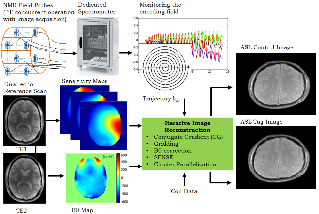
Signal Processing and Image Reconstruction: 16 k-space coefficients corresponding to a 3rd order spherical harmonic basis were computed from the probe signals for every time point during the imaging readout. Iterative conjugate gradient (CG)-based SENSE image reconstruction was performed on the coil raw data [5]. The comprehensive signal model used for image reconstruction is illustrated in Figure 1.
RESULTS
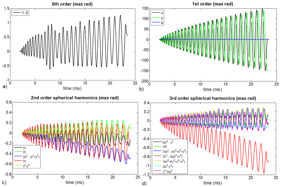
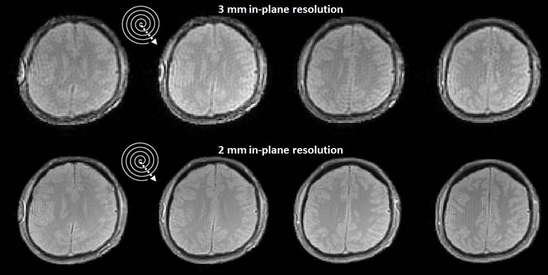
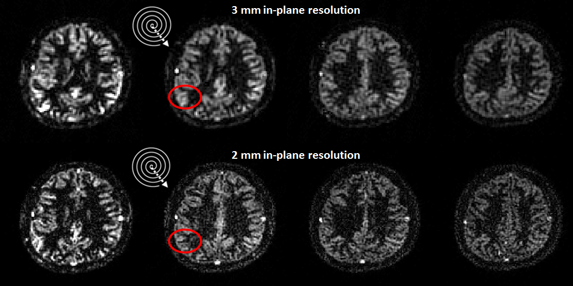
Figure 2 illustrates the monitored field evolutions up to 3rd order during spiral encoding. Figure 3 demonstrates the artifact-free ASL control images for 4 slices reconstructed from the single-shot spiral out scans combined with static off-resonance maps for 3 mm and 2 mm in-plane resolution. Corresponding ASL perfusion weighted images were calculated and averaged for 30 dynamics and plotted in Figure 4 exhibiting high gray and white matter contrast. Significant improvement in reducing the partial-volume effects was obtained with high SNR 2 mm in-plane resolution ASL perfusion images (red circles in Figure 4).DISCUSSION
Single-shot spiral ASL MRI in combination with image reconstruction based on a comprehensive signal model was presented. The reconstructed ASL images with an in-plane resolution of 3mm and 2 mm showed strongly reduced blurring- and aliasing artifacts. By applying field monitoring concurrently during all MR experiments, we also account for slow (quasi-static) B0 changes/drifts. At the same time, a consistent encoding geometry of the parametric input data such as ΔB0 maps and sensitivity maps is ensured. The TE of 3 ms corresponds to an increase of signal amplitude ~40% as compared to standard single-shot EPI (TE 18ms) for typical image resolutions in ASL perfusion weighted imaging of the brain. Besides increased SNR, shortened readout and TE durations with single-shot spiral acquisition particularly increase the motion robustness which is another important confounding in ASL-MRI [4].CONCLUSION
Single-shot spiral ASL with concurrent field monitoring largely overcomes the traditional vulnerability of spiral imaging in practice providing increased SNR, speed and motion robustness for research and clinical applications.Acknowledgements
No acknowledgement found.References
1. Glover GH. Spiral Imaging in fMRI. Neuroimage 2012;62:706–712.
2. King KF, Ganin A, Zhou XJ, Bernstein MA. Concomitant Gradient Field Effects in Spiral Scans. Magn. Reson. Med. 1999;41:103–112.
3. De Zanche N, Barmet C, Nordmeyer-Massner JA, Pruessmann KP. NMR Probes for Measuring Magnetic Fields and Field Dynamics in MR Systems. Magn. Reson. Med. 2008; 60:176–186.
4. Alsop DC, Detre JA, Golay X, Gunther M, Hendrikse J, Hernandez-Garcia L, Lu HZ, MacIntosh BJ, Parkes LM, Smits M, van Osch MJP, Wang DJJ, Wong EC, Zaharchuk G. Recommended Implementation of Arterial Spin-Labeled Perfusion MRI for Clinical Applications: A Consensus of the ISMRM Perfusion Study Group and the European Consortium for ASL in Dementia. Magn. Reson. Med. 2015;73(1):102-116.
5. Pruessmann KP, Weiger M, Börnert P, Boesiger P. Advances in Sensitivity Encoding with Arbitrary K-Space Trajectories. Magn. Reson. Med. 2001;46:638–651.
Figures