1020
Investigation of Simultaneous Multi-slice Imaging Utilized for Respiratory Gating: A Golden Angle Radial CAIPI Free-Breathing Cine MethodDana C Peters1, Chenxi Hu2, Yuqing Wan1, and Maolin Qiu1
1Yale University, New Haven, CT, United States, 2Yale University
Synopsis
We investigated simultaneous multi-slice as a method for performing respiratory-gating. One slice is used to monitor respiration, while another slice is targeted for imaging. Radial CAIPI data was acquired using a golden angle ordering for free-breathing 2D cine. At each cardiac phase and at each heart-beat, images of the heart and the diaphragm were obtained. Finally, a composite image of the heart was reconstructed using data from all frames acquired at end-expiration, with more projections and higher quality than a single heart-beat image.
Introduction
Here we
explore using “Controlled aliasing in parallel imaging results in higher
acceleration” (CAIPIRINHA, or CAIPI)(1) as a respiratory gating method. With two slices simultaneously excited, one
slice is used for respiratory monitoring while the other slice is the actual
target for imaging.
CAIPI acquires
multiple slices simultaneously, with different RF excitation phases for each
slice. The data is then reconstructed
using parallel imaging. Radial is a promising trajectory for CAIPI (2,3). Importantly, for retrospective respiratory-gating,
radial CAIPI with golden angles (4) allows flexible retrospective reconstruction,
generating a uniform distribution of k-space data. Methods
Methods: All imaging was performed on a 3.0T
Siemens scanner using a 32 channel cardiac coil (Invivo, Pewaukee, WI), in
subjects providing written informed consent. The overall acquisition scheme is
shown in Figure 1. CAIPI was implemented within a 2D GRE radial cine
sequence. To reduce eddy current effects
from rapidly changing spoke angles while also preserving the even coverage of
golden angle acquisition the cine data was acquired using a reordered golden
angle acquisition. A series of Npall
golden angles were
calculated. These angles were divided
into Nsets2π successive
sets which were reordered from 0 to 2π.
These segments were acquired in Npall /Nsets2π heart-beats, at multiple cardiac phases. ECG-gating and
free-breathing were used. The RF pulse was modified to excite two slices.
Within each heart-beat the RF excitation phase was alternated (0,π,0…) for the 2nd
slice. Two types of images were
generated (Figure 2). The
respiratory-slices were reconstructed with Np=Nsets2π projections,
to obtain an image of the diaphragm at any point chronological time during
scanning, and to monitor respiration. From these images, the heart-beats which
were timed to end-expiration were selected.
The target slice was then reconstructed using Np=Nsel· Nsets2π/2 data, where Nsel indicates the number of selected heart-beats timed to the end-expiratory phase, and only the first half (0-π), or 2nd half (π -2π) of the projections in a single segment were used, for
a reduced acquisition window. All radial CAIPI data was reconstructed with parallel imaging
using Algebraic Reconstruction Technique (ART) (6) (9 iterations, λ=0.1), an iterative method similar to
projection onto convex sets (POCS). The
coil-maps estimates were generated from all acquired projections. Typical scan parameters were: TR/TE/θ=5.5ms/2.0ms/12◦, FOV=36cm, Hz/pix=400Hz,
slice=10mm, slice gap ~ 10cm, Npall=512, Nsets2π=32, 88ms per phase.
Results
Figure 3 shows phantom data using golden angle radial CAIPI, scanned with a 32 channel head coil, showing reasonable quality. Figure 4 shows an undersampled respiratory slice (32 Np), the target slice (16Np) from a single heart-beat, the target-slice from all heart-beats (which is blurry), and finally the targeted slice reconstructed with selected projection sets (here using 4 end-expiratory heart-beats, i.e. 64 Np). This data is from a healthy volunteer.Discussion
We present a novel method for CAIPI respiratory gating, which uses CAIPI to excite simultaneously one slice for monitoring respiration, and another for imaging. We demonstrate its feasibility in initial experiments. Respiratory compensation is still a key challenge in cardiac imaging, especially for high resolution applications, and for poor breath-holders. Many novel solutions exist beyond traditional breath-holding, bellows-gating, and navigator-gating: e.g. self-gating and image-based gating (6-8) methods, among others. CAIPI-gating provides an almost-ideal respiratory monitoring technique.Acknowledgements
No acknowledgement found.References
1) Beuer FA et al., MRM 2005. 2) Wang H, et al., MRI 2016. 3) Yutzy SR et al., MRM 2011. 4) Winkelman S et al., IEEE TMI 2007. 5) Li S et al. MRM 2014. 6)Larson AC et al., MRM 2005. 7) Stehning C et al., MRM 2005. 8) Henningsson M et al., MRM 2015.Figures
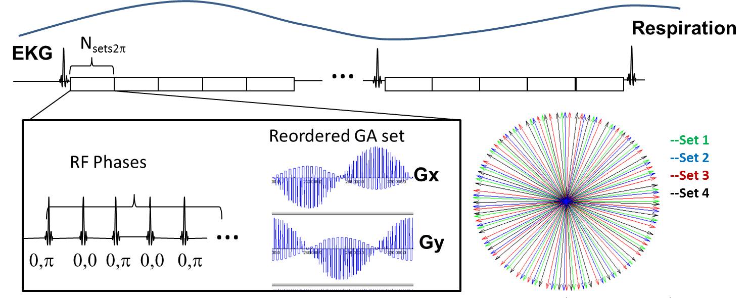
Figure 1: Free-breathing ECG-gated GRE
cine data were acquired using radial CAIPI. Two slices were acquired
simultaneously, with RF phase-cycling for the 2nd slice. Golden angle ordering
was used, with the subset of angles (Nsets2π)
within each cardiac phase sequentially reordered to span 0 to 2π. In
subsequent heart-beats additional sets of angles were acquired.
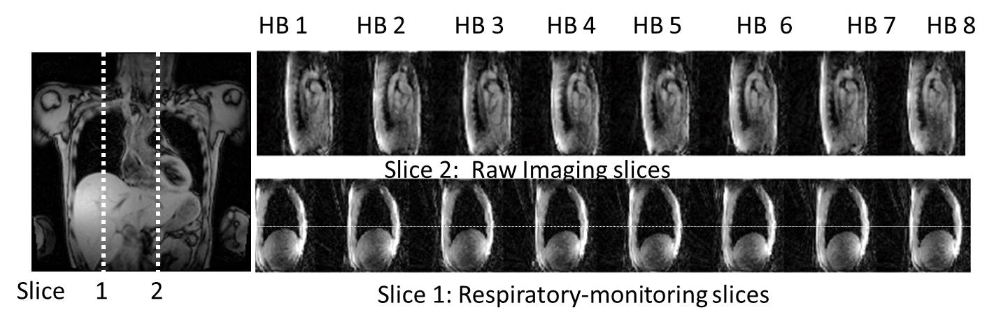
Figure 2: In CAIPI-gated imaging, two slices are excited. One slice monitors the diaphragm, generating an image at any point during the acquisition. The other slice is the target slice, which is acquired with successive sets of projections in each heart-beat, using a golden angle acquisition order. The CAIPI-gating data is then used to select the heart-beats (e.g. end-expiratory) which will be included in a final composite reconstruction.
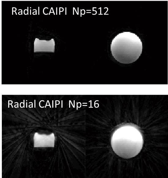
Figure 3: Phantom study using golden
angle radial CAIPI, reconstructed with 512 and 16 Np, using parallel imaging.
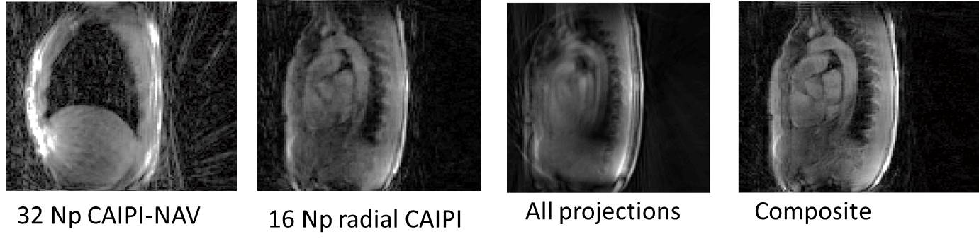
Figure 4: Images from a healthy subject
showing the diaphragmatic slice and the target slice reconstructed with data
from one heart-beat, and the target slice reconstructed using all of the
data. The composite image reconstructed from selected heart-beats at
end-expiration shows improved quality.