0978
Accelerated 3D bSSFP Imaging for Treatment Planning on an MRI-Guided Radiotherapy System1Radiology, University of California, Los Angeles, Los Angeles, CA, United States, 2Physics and Biology in Medicine IDP, University of California, Los Angeles, Los Angeles, CA, United States, 3Radiation Oncology, University of California, Los Angeles, Los Angeles, CA, United States
Synopsis
The purpose of this work is to introduce a compressed sensing and parallel imaging combined technique to reduce the acquisition time for planning MR. We implemented a variable-density Poisson-Disk under-sampled acquisition along with L1-ESPIRiT reconstruction technique on an MRI-guided radiotherapy system. Phantom study showed that our technique had superior image quality over the conventional GRAPPA approach. Patient and volunteer study demonstrated that comparable images can be acquired with half of the original time. In addition, the proposed technique was able to achieve
Purpose:
MRI-guided radiotherapy (RT) system is gaining momentum in the field of radiation oncology benefited from the superior soft tissue contrast of MR for organ delineation and motion tracking1-4. Organ delineation and treatment planning primarily based on MR images from the MRI-guided RT system (MRIdian, ViewRay, Oakwood Village, OH) is the current standard in our institute for patients treated on ViewRay. However, unlike the “snap-shot” CT imaging, acquisition time of MR is relatively long, so that breath-hold is often required for abdominal cancer patient to avoid breathing motion deteriorating the image quality. This, in the meantime, leads to a tradeoff between spatial coverage, spatial resolution, image quality, and scan time. The current protocol used in our institute required a 25s breath-hold, which is relatively long for some patients. The purpose of this work is to introduce a compressed sensing and parallel imaging combined technique, which is universally applicable to all MRI-guided RT system, to greatly reduce the acquisition time and increase patient comfort. Gained time can be used for higher resolution imaging that would benefit the subsequent treatment planning workflow.Methods:
Data Acquisition and image reconstruction: The original planning MR is based on the balanced steady state free precession(bSSFP) sequence with an effective under-sampling rate of 7.5 fold (generalized autocalibrating partially parallel acquisitions (GRAPPA) 2×2, elliptical scanning, and 6/8 partial Fourier in both phase and slice directions). To achieve a higher under-sampling rate without suffering from severe artifacts and noise amplification associated with GRAPPA acquisition, ky-kz encoding plane was modified in a way that the center 22×16 region was fully sampled while the outer region followed a variable-density Poisson-Disk distribution5. Two acceleration rates: 7.5 fold(7.5x) and 15 fold(15x) were programed. All data were acquired on the 0.35T MR scanner of the ViewRay system. L1-ESPIRiT algorithm6 was used to reconstruct the under-sampled Poisson-Disk data: $$x=argmin\sum_{i=1}^{N}||DFS_ix-m_i||_2^2+\lambda||Wx||_1$$
where $$$D, F, S_i, x, m_i, \lambda, W$$$ are the data selection operator, the Fourier transform, estimated sensitivity maps, image to be reconstructed, acquired under-sampled data from coil $$$i$$$, regularization weighting, and Daubechies wavelets respectively.
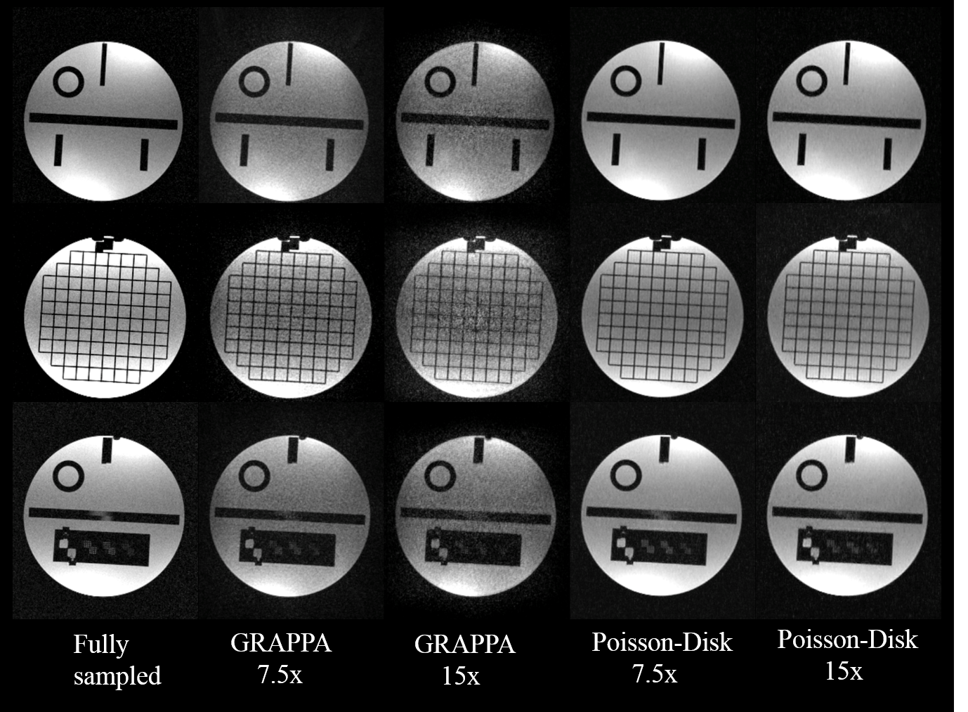
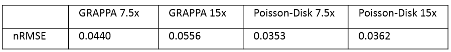
Phantom Study: Phantom validation was performed on the standard American College of Radiology phantom. One fully-sampled and four under-sampled (using GRAPPA and variable-density Poisson-Disk with 7.5x and 15x acceleration rate respectively) data were acquired. Normalized root-mean-square errors (nRMSE) was calculated for all slices between the fully-sampled images and under-sampled images.
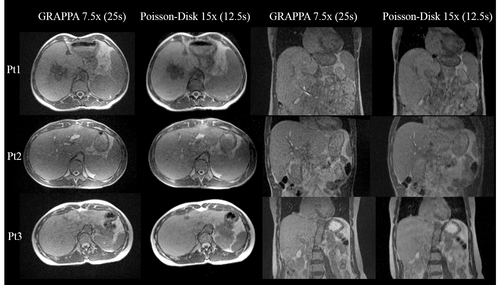
In-vivo Study: Five patients and three volunteers were included in the study. Two breath-hold images were acquired using GRAPPA 7.5x (25s) and Poisson-Disk 15x (12.5s)(resolution=1.5×1.5×2 mm3, coverage=500×449×228mm3). Among all subjects that successfully performed the breath-hold, liver boundary sharpness was measured by drawing a line across the boundary in the coronal plane, where the sharpness is defined as the inverse of the distance between the two points having 20% and 80% signal intensity among the dynamic range. Paired t-test was used to assess whether there is a significant difference between the two techniques in terms of sharpness. One 1×1×1mm3 isotropic high-resolution study was conducted on one volunteer to verify the feasibility of achieving high-resolution imaging.
Results and Discussion:
The reconstruction was implemented based on the Berkeley Advanced Reconstruction Toolbox7,8(~1 minute per 3D volume). Figure 1 shows the phantom results on three representative slices. With the increase of under-sampling rate, noise amplification is apparent in GRAPPA accelerated images, whereas negligible in Poisson-Disk accelerated images. This is further supported by the quantitative nRMSE calculation in Table 1.
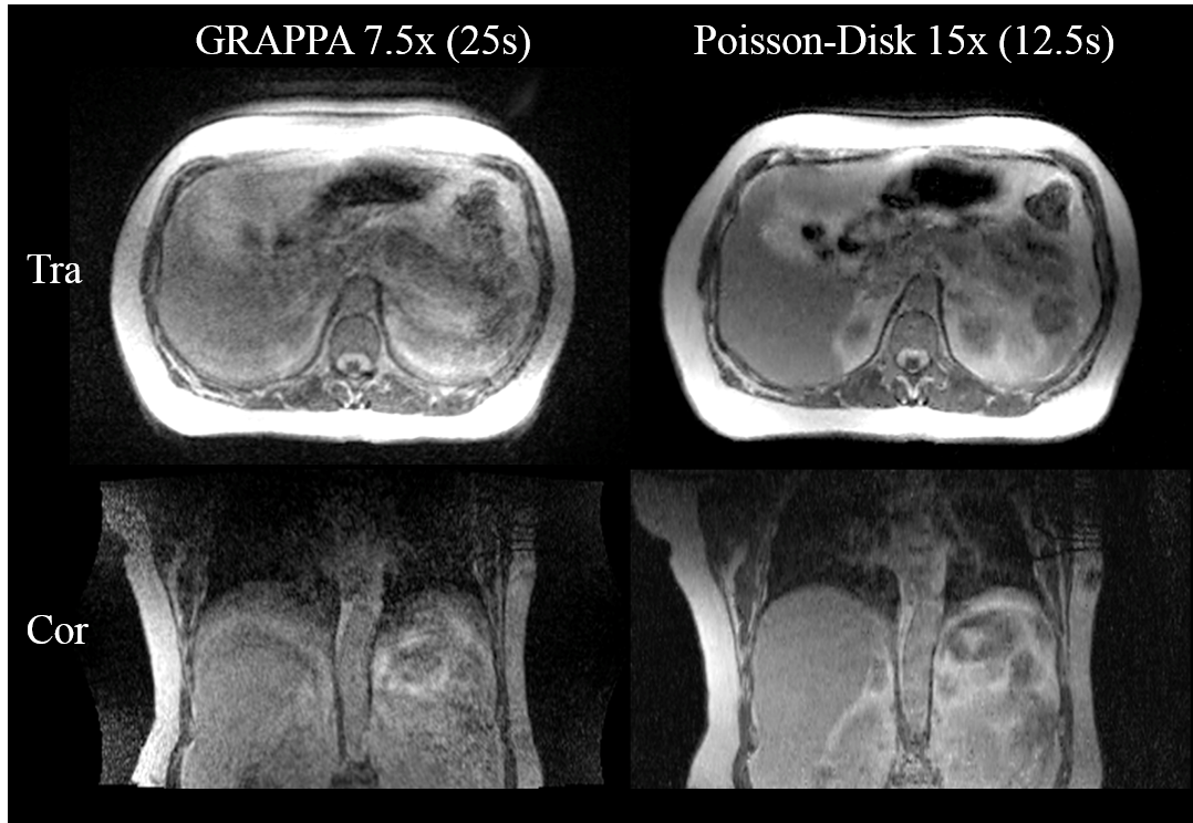
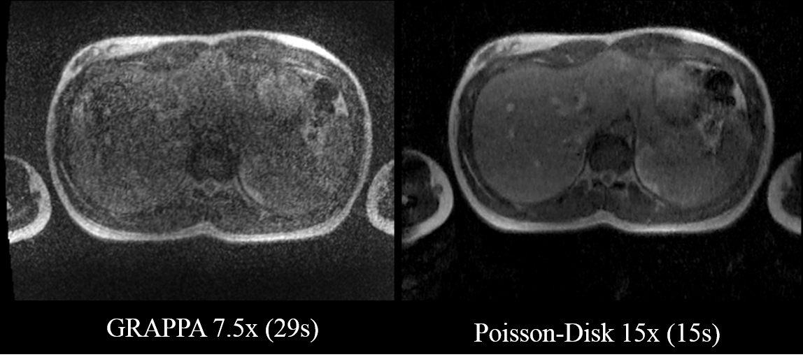
Breath-hold was successfully performed in seven out of eight subjects. The mean boundary sharpness calculated from GRAPPA accelerated images and Poisson-Disk accelerated images were 0.374±0.073(mm-1) and 0.361±0.075(mm-1) with no statistically significant difference (p=0.429). Figure 2 shows the comparison between the 25s GRAPPA technique and 12.5s Poisson-Disk technique on three liver patients where all breath-holds were successfully performed. Our proposed method provided comparable image quality with only half of the scan time. Figure 3 is an example where the patient was not able to hold the breath for 25s whereas the 12.5s breath-hold was more feasible for her. Under high-resolution scenario as shown in Figure 4, Poisson-Disk technique can still maintain desirable image quality while noise level is unacceptably high in GRAPPA accelerated images, implying that GRAPPA is infeasible to be used for high-resolution imaging on this low field strength system.
Conclusion:
This proof-of-principle study validated the feasibility of using variable-density Poisson-Disk sampling pattern along with L1-ESPIRiT reconstruction technique to accelerate the planning MR acquisition on a 0.35T MR-guided RT system. The proposed technique could provide sufficient coverage, desirable image quality, shorter acquisition time and potentially higher resolution imaging for treatment planning.Acknowledgements
Yu Gao acknowledges research support from ViewRay.References
[1] S. Mutic and J. F. Dempsey, “The ViewRay System: Magnetic Resonance–Guided and Controlled Radiotherapy,” Semin. Radiat. Oncol., vol. 24, no. 3, pp. 196–199, Jul. 2014.
[2] B. W. Raaymakers et al., “Integrating a 1.5 T MRI scanner with a 6 MV accelerator: proof of concept,” Phys. Med. Biol., vol. 54, no. 12, p. N229, 2009.
[3] P. J. Keall, M. Barton, and S. Crozier, “The Australian Magnetic Resonance Imaging–Linac Program,” Semin. Radiat. Oncol., vol. 24, no. 3, pp. 203–206, Jul. 2014.
[4] “Products – MagnetTx.” [Online]. Available: http://www.magnettx.com/index.php/category/products/. [Accessed: 12-Oct-2016].
[5] R. L. Cook, “Stochastic Sampling in Computer Graphics,” ACM Trans Graph, vol. 5, no. 1, pp. 51–72, Jan. 1986.
[6] M. Uecker et al., “ESPIRiT-an eigenvalue approach to autocalibrating parallel MRI: Where SENSE meets GRAPPA,” Magn. Reson. Med., vol. 71, no. 3, pp. 990–1001, Mar. 2014.
[7] M. Uecker et al., "Software toolbox and programming library for compressed sensing and parallel imaging," In Proceedings of the ISMRM Workshop on Data Sampling and Reconstruction, Sedona, Arizona, USA, 2013.
[8] BART: version 0.2.04 (2014) DOI: 10.5281/zenodo.12495.
Figures