0971
Multi-slice metabolite mapping with Very-High Degree Dynamic B0 Shim Updating at 9.4T using Accelerated 1H FID MRSI1MPI for Biological Cybernetics, Tuebingen, Germany, 2IMPRS for Cognitive and Systems Neuroscience, Eberhard Karls University of Tübingen, Tuebingen, Germany, 3Institute of Physics, Ernst-Moritz-Arndt University Greifswald, Greifswald, Germany
Synopsis
In this work, we address the problem of B0 inhomogeneity in the human brain at 9.4T by using dynamic very high order B0 shimming. This enables multi-slice metabolite mapping in the human brain at this field strength. Furthermore, we investigate the advantage of low (2nd) versus very high (4th+) degree dynamic B0 shimming directly with respect to the quality of the metabolite maps.
Purpose
Demonstrate the advantage of very-high degree dynamic B0
shimming for multi-slice metabolite mapping at 9.4T.Introduction
The increased SNR of ultra-high field (UHF) strengths is beneficial to MRSI. It allows us to go to higher spatial resolutions. Despite the advantages of going to higher field strengths, there are still many challenges such as B0 and B1 inhomogeneity. The B0 inhomogeneity, in particular, can cause a loss of SNR, signal dropout and broader spectral linewidths. It has previously been shown that very-high degree static B0 shimming as well as lower degree slice-wise dynamic shim updating can positively affect the linewidths for MRSI1-6.
In this work, we thus address the problem of B0 inhomogeneity in the human brain at 9.4T by using dynamic high-order B0 shimming. This enables multi-slice metabolite mapping in the human brain at this field strength. Furthermore, we investigate the advantage of low (2nd) versus very high (4th+) degree dynamic B0 shimming directly with respect to the quality of the metabolite maps.
Methods
A Siemens 9.4T whole-body MRI scanner with an in-house 8-channel transceiver radiofrequency coil7 was used to acquire the data. A 28-channel insert shim, with up to 4th degree and partial 5th degree shim terms (4th+ degree), from Resonance Research Inc. (Billerica, MA) was used for dynamic B0 shimming. The insert shim was calibrated by modelling the actual shim fields2. The optimal slice-wise B0 shim was calculated using a constrained regularised inversion8.
The dynamic B0 shimming was performed using a custom MatlabTM script to update the shim amplifiers via the RS232 interface of the respective 28-channel amplifiers (RS232 was converted to/from RS422 for robustness). An external TTL trigger from the MRI sequence was sent at the beginning of each slice acquisition to signal that the shim values needed to be updated.
High resolution MRSI data were acquired using a 2D multi-slice FID MRSI sequence9 with a R = 2x2 SENSE acceleration10. The parameters were: FOV 200mmx200mm, matrix size 64x64x3, slice thickness 10mm, distance factor 100%, TR 220ms, acquisition delay 1.5ms. The total volume coverage was 5cm. An optimised three-pulse water suppression was used9. The acquisition was repeated with both low (2nd) and very high degree (4th+) dynamic B0 shimming. The spectra were fit using LCMODEL and metabolites were mapped and compared for both shim settings.
Results
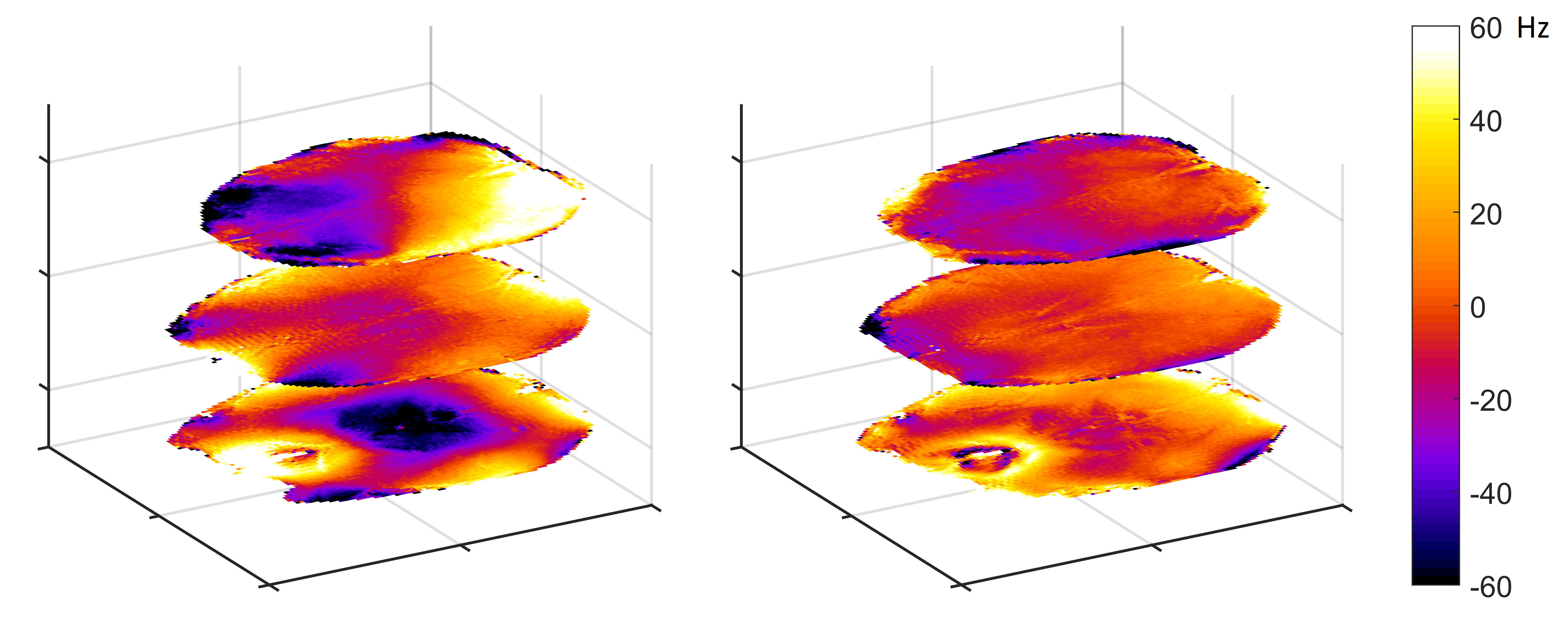
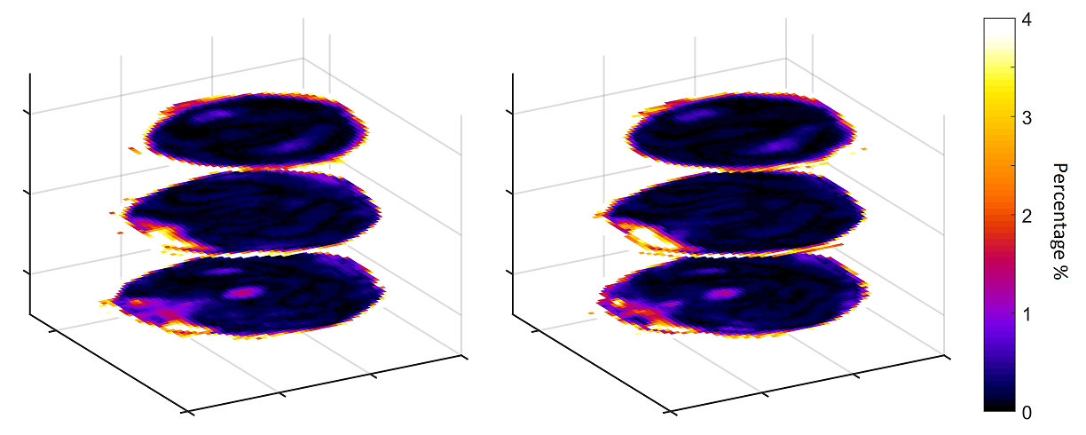
The bottom slice in this study was chosen low enough to be affected by the nasal cavity. The shimmed B0 maps resulting from dynamic shimming using 2nd and 4th+ degree shimming are shown in Fig. 1. The maps show the advantage of higher degree B0 shimming, especially for the top and bottom slices. As a quality control measure, Fig. 2 shows the water suppression maps. The maps show that the water suppression is robust against B0 and B1 inhomogeneity and the water suppression factor is less than 1% for most voxels.
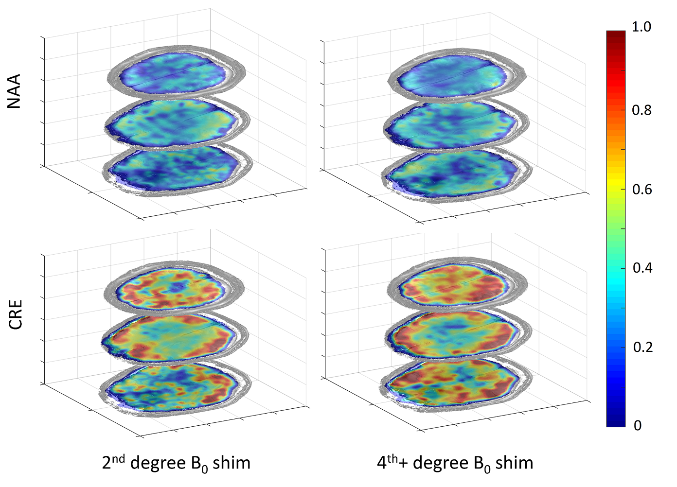
Metabolite maps are shown for NAA and Creatine in Fig. 3. It can be seen that some spatial variations in the maps of the 2nd degree shimming do not correspond to anatomy and are instead an artifact of unreliable fitting. However, the 4th+ degree shimming maps show better white/gray matter contrast especially for Cre that reflect the underlying anatomy more accurately. Furthermore, we can see from the full-width half-maximum (FWHM) and CRLBs of Creatine (in Fig. 4) that the linewidths are narrower using slice wise dynamic 4th+ degree B0 shimming and thus the confidence of the fit is also better.
Discussion
The results show that dynamic B0 shimming can still benefit from higher degree shim terms. 2nd degree shim terms, even when slice wise dynamically updated, are insufficient for reliable multi-slice metabolite mapping and quantification in the human brain at 9.4T. A drawback of the current very high degree insert shim is that more eddy currents are induced in the shim coils (especially for gradient switching intensive scans). More eddy currents result in phase problems and could cause fitting errors. Furthermore, the 4x acceleration used to enable shorter scan times comes at the price of reduced SNR.
Conclusion
For the first time at 9.4T the advantage of very-high degree dynamic slice wise B0 shim updating was shown which enabled multi-slice metabolite mapping by FID MRSI. Interestingly, the B0 homogeneity not only affects the confidence of the fit but one can clearly see visible changes in the metabolite concentration maps. Therefore, if the B0 shim is poor, differences between metabolite concentrations can result from differences in B0 inhomogeneity (for spectroscopy-imaging and single-voxel spectroscopy) and not because of the underlying anatomy.Acknowledgements
This study was supported by the European Research Council Starting grant, project SYNAPLAST MR #679927.References
[1] Hetherington H et al. MRM. 2006. [2] Chang P et al, ISMRM spectroscopy workshop 2016. [4] Pan J et al, MRM 2012. [5] Fillmer A et al, MRM 2015. [6] Juchem C et al, Concepts Magn. Res. B 2010. [7] Avdievich N et al. NMR in Biomed 2016. [8] Nassirpour S et al. Proc of ISMRM 2016. [9] Nassirpour S et al., ISMRM spectroscopy workshop 2016. [10] Dydak U et al, MRM 2001.Figures