0967
An integrated 32ch RF-shim array coil for improved B0 shimming of the brain at 7 Tesla1A. A. Martinos Center for Biomedical Imaging, Massachusetts General Hospital, Charlestown, MA, United States, 2Harvard Medical School, Boston, MA, United States, 3Electrical Engineering and Computer Science, Massachusetts Institute of Technology, Cambridge, MA, 4Institute of Biomedical Engineering, National Taiwan University, Taiwan, 5Electronic and Mechatronic Systems, Technische Hochschule Nürnberg, Nürnberg, Germany, 6Harvard Medical School, Boston, United States, 7Electrical Engineering and Computer Science, Massachusetts Institute of Technology, Cambridge, MA, United States
Synopsis
Dual-purpose arrays of close-fitting coils, used to both receive RF and shim B0 to high spatial order, have recently been demonstrated in brain imaging at 3T. We extend this approach to 7T with an array comprised of: six RF coils, twenty-six RF/B0-shim coils, and six shim-only coils. The shim coils are driven by an array of low-cost current-feedback amplifiers that can steer up to fifty amps of shimming currents rapidly and accurately enough to allow for slice-by-slice adjustments. The resulting slice-optimal high-order shim capability (wires and chokes) improves σB0 in brain slices by up to 60% (as compared to static 2nd-order global shimming), while worsening SNR by less than 10%, and increasing coil coupling by less than 3%.
Motivation
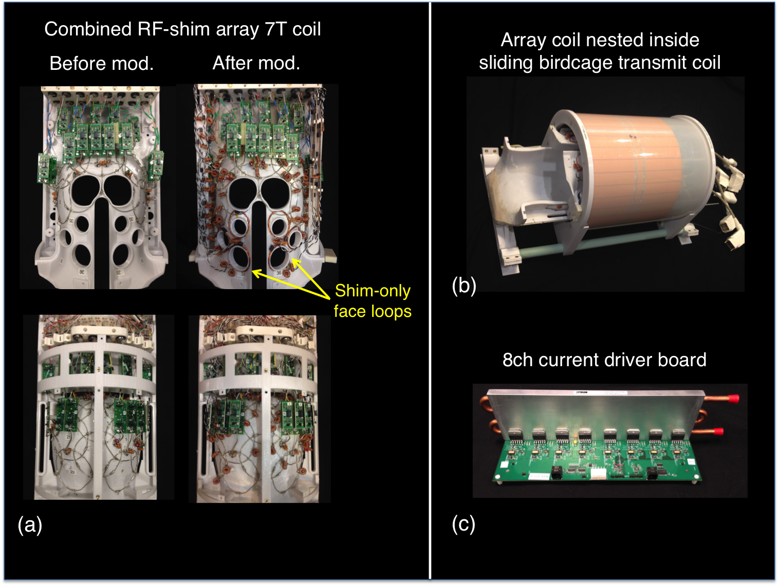
Multi-coil (MC) shimming [1] has been proposed as an alternative or complement to spherical harmonic (SH) shim coils for nulling static and dynamic high-spatial order B0 inhomogeneity in the brain. MC arrays of small loops patterned close to the body can be driven by low-cost current sources and switched rapidly for dynamic shimming due to their low inductance and minimal coupling to conducting structures in the bore. Recently, MC shimming has been realized using integrated 3T RF-shim arrays, allowing both types of coils to occupy space close to the body where they function with greatest efficiency, rather than compete for space [2, 3]. We extend this approach to brain imaging at 7T, where spatiotemporal variations in ΔB0 are more severe. In our “hybrid” 32ch design, 6 channels use RF-only loops and 26 channels use combined RF-shim “AC-DC” loops. Additionally, 6 shim-only loops are placed over the face for targeted shimming of the frontal lobe [4, 5]. We describe the design of the RF-shim coil and assess its RF and B0 shimming performance in vivo.Methods
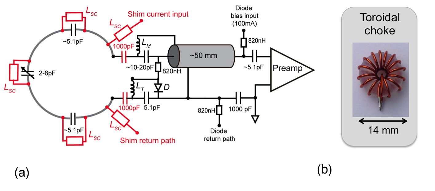
Figure 1 shows the conventional 32ch 7T RF receive-array paired with a detunable birdcage transmit coil. The SNR performance was tested prior to adding shim components. Figure 2 shows the RF coil circuit along with the chokes needed to bridge shim current into the loop and across distributed tuning capacitors. The chokes are self-shielding toroidal inductors (15-turn, AWG22, 13.5mm O.D., 4mm I.D.). They have self-resonance near 320MHz and RF impedance > 2kΩ at the Larmor frequency (297MHz). After being added to a coil, choke windings are hand-tuned to eliminate any parasitic resonances and ensure high isolation between the RF coil and the DC feed wires. To limit coupling between the transmit coil and the feed wires, chokes are placed at >10cm intervals along each wire. The B0 profile of each shim coil is field mapped to create a basis set for in vivo shimming. RF SNR maps [6] are acquired on an anthropomorphic head phantom [7] before and after adding shim hardware. A 90° flip-angle is used to reduce the effect of B1+ variations on signal intensity. Safety tests are performed for RF power absorption and gradient-induced eddy-current heating [8]. In vivo shimming is performed on one healthy volunteer. The brain is shimmed up to 2nd-order using conventional system SH shims then baseline ΔB0 field maps are acquired. Optimal MC shim currents are calculated using MATLAB's 'fmincon' (limit: 3A/ch or 50A total), and then MC shim currents are applied. The shim coils are driven by digitally-programmable, open-source current drivers (Figure 1) [9]. Shim performance is evaluated using the standard deviation of the ΔB0 field maps (σB0) and distortion in unaccelerated single-shot EPI axial slices acquired with both P-A (“blip-up”) and A-P (“blip-down”) phase encoding directions.Results
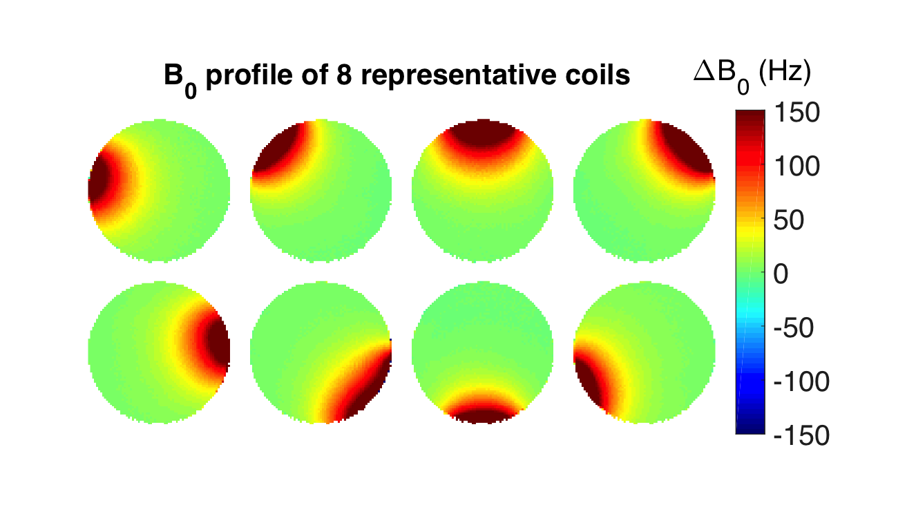
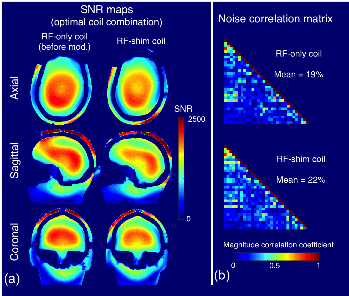
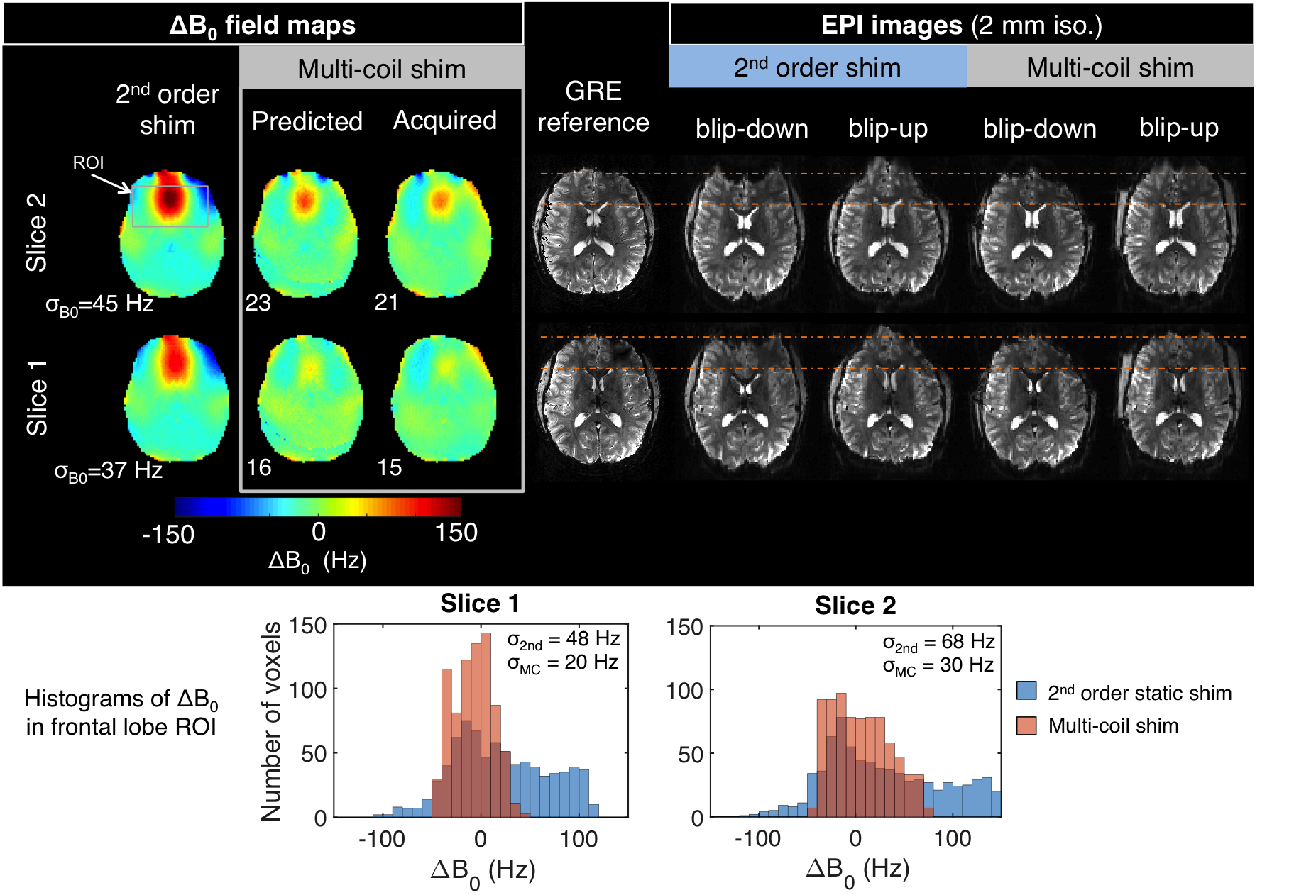
The current driver boards deliver stable currents to each coil and do not introduce structured image artifacts or RF noise. Figure 3 displays ΔB0 field maps from 8 representative coils, showing their spatially-distinct field profiles. Differences in the birdcage coil transmit adjust reference voltage before and after coil conversion were within +/-10% (similar to normal day-to-day variation). Figure 4 shows that the shim hardware causes an approximately 5-10% decrease in average SNR and 3% increase in coil-coil coupling coefficient. MC shimming of a 1cm slab decreases σB0 by 53% and 60% in two slices as compared to static 2nd-order shims and narrows the histogram of ΔB0 values in a fontal lobe ROI (Figure 5). Distortion in 2mm iso. EPI slices is visibly reduced compared to an undistorted image.Discussion
Because safety testing was only performed after shim hardware was added, in vivo SNR maps could not be obtained for the RF-only configuration. The head phantom used for SNR mapping shows pronounced central-brightening due to dielectric wavelength effects that causes the SNR profile to differ from profiles observed in vivo. However the SNR maps are still considered a useful measure of the relative SNR of the two coil configurations, since differences in the signal intensity arise from changes in the coil receive sensitivity rather than changes in flip angle.
We demonstrate that MC shimming hardware can be safely integrated into a 7T RF receive-array without compromising transmit efficiency and with a relatively modest impact on receive coil performance (SNR and noise correlation matrix). Proof-of-concept MC slab shimming reduces σB0 in brain slices by 50% or more compared to 2nd-order shims. In future work, the array coil holds promise for dynamic, slice-optimized shimming and real-time compensation of B0 fluctuations arising from physiological processes.
Acknowledgements
The authors thank Simon Sigalovsky for assistance with mechanical fabrication. Grant support comes from NIH K99 EB021349, NIH R21 EB017338, and NIH P41 EB015896.References
[1] Juchem C, Nixon TW, McIntyre S, Boer VO, Rothman DL, and de Graaf RA, Dynamic Multi-Coil Shimming of the Human Brain at 7 Tesla. J Mag Reson, 2010;212(2):280-288.
[2] Truong TK, Darnell D, Song AW. Integrated RF/shim coil array for parallel reception and localized B0 shimming in the human brain. NeuroImage 2014;103:235-40
[3] Stockmann JP, Witzel T, Keil B, Polimeni JR, Mareyam A, LaPierre C, Setsompop K, Wald LL. A 32-channel combined RF and B0 shim array for 3T brain imaging. Magn Reson Med. 2016;75(1):441-51.
[4] Juchem C, Nixon TW, McIntyre S, Rothman DL, de Graaf RA. Magnetic field homogenization of the human prefrontal cortex with a set of localized electrical coils. Magn Reson Med. 2010;63(1):171-80. et al., MRM 2010.
[5] Stockmann JP, Guerin B, Wald LL. Improving the efficiency of integrated RF-shim arrays using hybrid coil designs and channel placement and compression via a genetic algorithm. ISMRM 2016, p. 1153.
[6] Kellman P and McVeigh ER. Image reconstruction in SNR units: a general method for SNR measurement. Magn Reson Med. 2005;54(6):1439-47.
[7] Guerin B, Stockmann JP, Baboli M, Torrado-Carvajal A, Stenger AV, Wald LL. Robust time-shifted spoke pulse design in the presence of large B0 variations with simultaneous reduction of through-plane dephasing, B1+ effects, and the specific absorption rate using parallel transmission. Magn Reson Med. 2016;76(2):540-54.
[8] Keil B, Blau JN, Biber S, Hoecht P, Tountcheva V, Setsompop K, Triantafyllou C, and Wald LL. A 64-channel 3T array coil for accelerated brain MRI. Magn Reson Med. 2013;70(1):248-58.
[9] Arango N, Stockmann JP, Witzel T, Wald LL. Open-source, low-cost, flexible, current feedback-controlled driver circuit for local B0 shim coils and other applications. ISMRM 2016, p. 1157.
Figures