0944
Correlation between Breast Cancer Molecular Subtypes and Parameters of Dynamic Contrast Enhanced (DCE) MRI and Intravoxal Incoherent Motion (IVIM): Implication for Breast Cancer Anti-angiogenesis Treatment Guidance1Radiology, Koo Foundation Sun Yat-Sen Cancer Center, Taipei, Taiwan, 2Research, Koo Foundation Sun Yat-Sen Cancer Center, Taipei, Taiwan
Synopsis
This prospective study correlates DCE MRI and IVIM with breast cancer molecular subtypes by examine the differences in vascular normalization signature genes. We found molecular subtype III and VI with higher pericyte gene scores to have significantly lower perfusion related parameters, higher extracellular extra-vascular space on DCE MRI and IVIM. These associations may be used to guide anti-angiogenesis treatment for breast cancer.
Purpose:
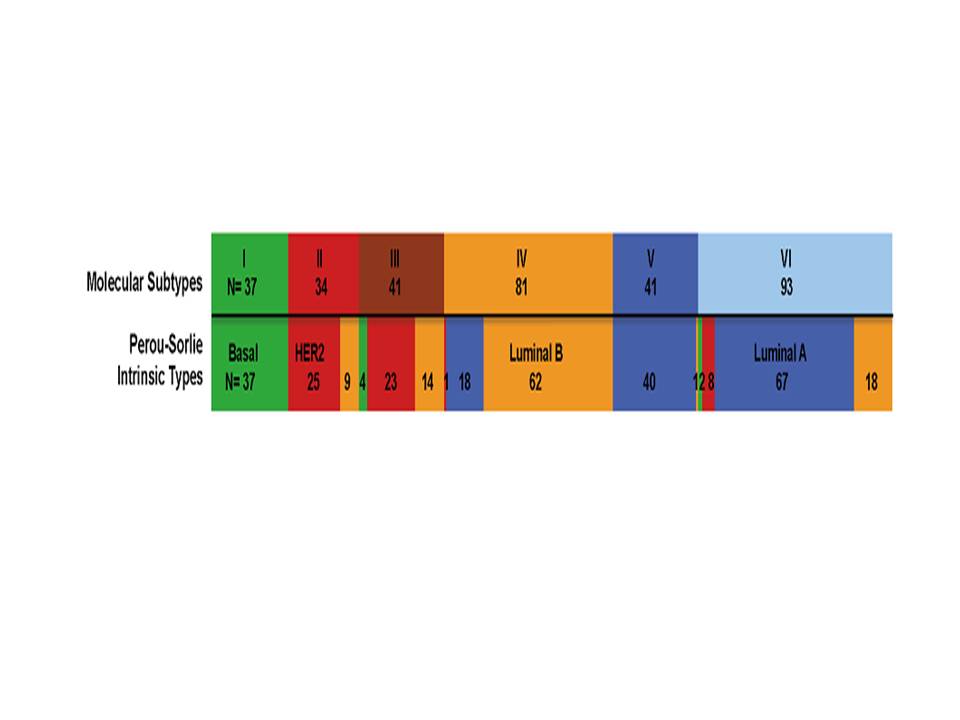
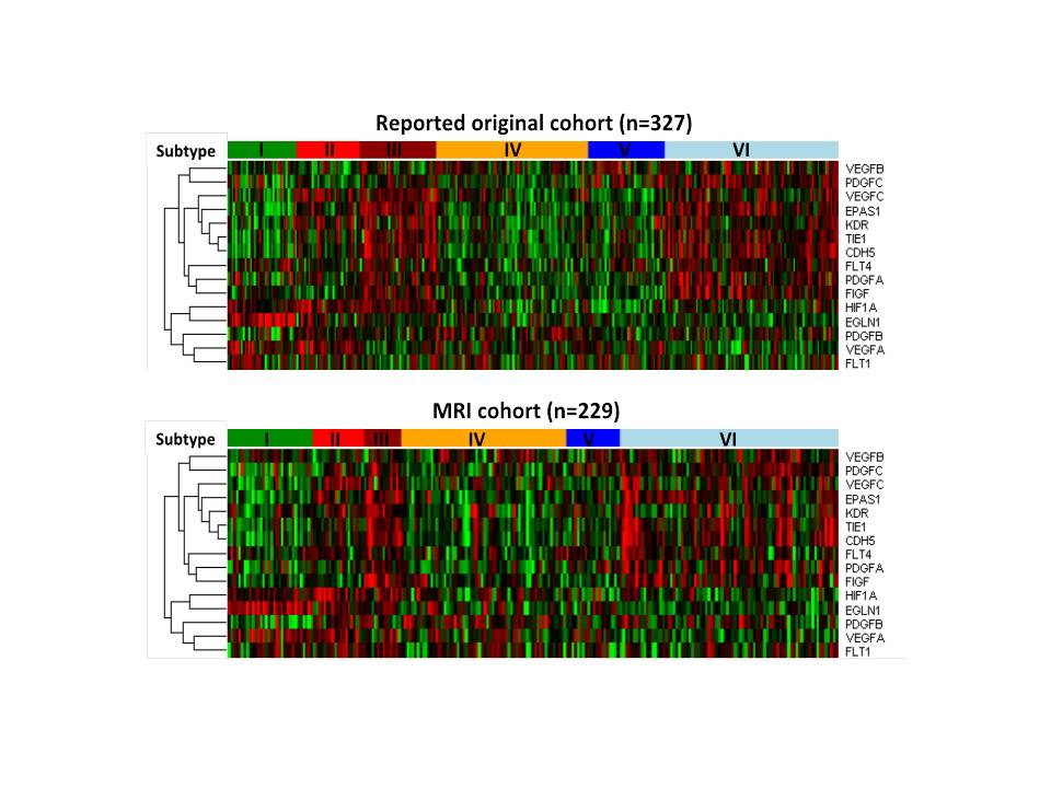
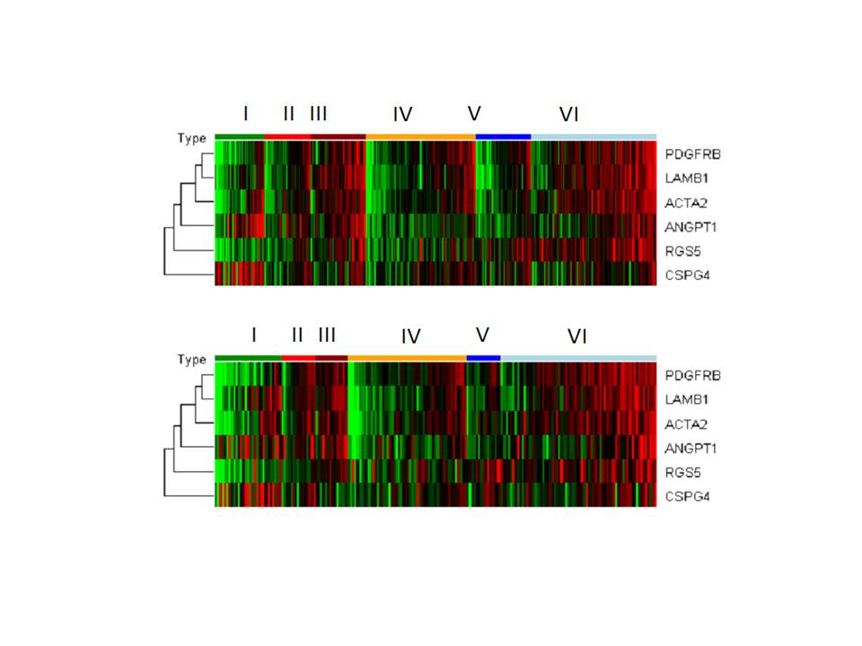
Six different molecular subtypes in breast cancer were established in an earlier study we did on expression of 783 genes 1 (Figure 1). A subsequent study further revealed that subtypes III and VI had increased expression of vascular normalization and pericyte signature genes 1 2(Figures 2 and 3 upper panel), which suggested the features of more normal vascular structure and less leakiness in these two subtypes of breast cancer. The purpose of this current study is to determine whether vascular function measured by dynamic contrast enhanced (DCE) magnetic resonance imaging (MRI) and intravoxel incoherent motion (IVIM) can be correlated with the molecular subtypes.Material and Methods:
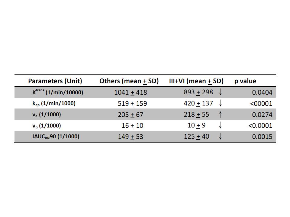
With IRB approval and signed informed consent, 229 de novo breast cancer patients with tumor sizes between 1.5 and 5 cm were recruited into the study. DCE breast MRI was performed before surgery. Breast MRI was conducted on a 1.5T scanner (Optima MR450W, GE, WU) using the body coil as a transmitter and an 8-channel biltateral phased array breast coil as signal reception. The imaging position was centered on the tumor, with a 6-cm slab thickness for the following sequences: Pre-contrast T1 map and B1 map with matched slices and resolution acquired for T1 correction. T1 map were acquired using SPGR sequence with five flip angles (FAs) from 2° to 30°. Axial 3D T1W fat-saturated DCE-MRI acquired by volume image breast assessment (VIBRANT) gradient echo sequence with the following parameters: TR/TE=3.8 ms/1.2ms; FA= 12; BW= 41.67 kHz; FOV= 34×34 cm; matrix size= 96×80; and slice thickness= 3 mm for 20 continuous slices per phase with a temporal resolution of 4.49 seconds. (Total 75 phases: 5 pre and 70 post IV contrast) IVIM DWI studies were performed on the following parameters: TR/TE=8000 ms/65.9ms; FOV= 34×34 cm; matrix size= 98×128; slice thickness= 5 mm; slice gap= 0.5 mm; NEX= 3; The diffusion gradient encoding in three orthogonal directions was used with 11 b values (from 0 to 1200 s/mm2) covering the whole breast. The pharmacokinetic parameters were estimated by fitting two-compartment Tofts model3. Segmented biexponential IVIM analysis was performed to estimate Dt, f, and Dp4. A single operator drew ROIs around the outer tumor border limiting DCE MRI and IVIM analysis to the maximum axial tumor area section. Gene expression profiling by Affymetrix U133 plus2.0 array and molecular subtype determination was conducted on fresh biopsy or frozen surgical breast cancer tissue 1. Expression of six genes (ACTA2, ANGPT1, CSPG4, LAMB1, PDGFRB and RGS5) associated with pericytes was measured for hierarchical-clustering and principal components analysis. The concordance of different gene expression for six molecular subtypes between our previous studied cohort (n=327) and this MRI cohort (n=229) was analyzed by Pearson correlation using permutation test (10000 permutations). When comparing pericyte gene scores in among different molecular subtypes, Mann-Whitney test with Bonferroni correction with p value < 0.05/6 ≈0.0083 indicate significant higher in subtype III and VI than others. When comparing DCE-MRI and IVIM parameters mean of subtype III and VI with others, Mann-Whitney test was used at significance level of 0.05.Results:
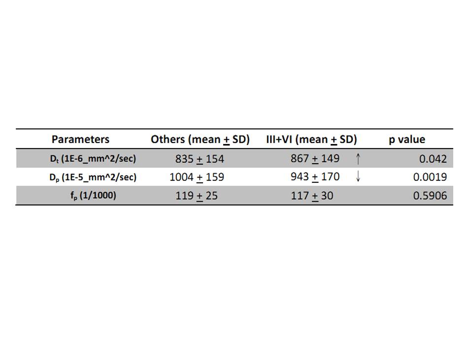
We examined all the breast cancer patients (n=229) for gene expression of vascular normalization and pericyte signatures by subtypes. The results mirrored our earlier findings that only subtypes III and VI (n=96) had increased expression of both signatures (concordance between two cohorts: Pearson correlation r=0.89, p<0.0001. Figure 2, 3 lower panel). The pericyte scores in subtype III and VI are significantly higher than those in other subtypes (p=0.0024 and p<0.0001, respectively). The comparisons in parameters of DCE MRI and IVIM study among the different subtypes of breast cancers showed that molecular subtypes III and VI breast cancers had significantly lower mean perfusion related parameters: lower Ktrans (p=0.0404), kep (p<0.0001), vp(p<0.0001), IAUGCBN90 (p=0.0015), Dp(p=0.0019); higher extravascular extracellular space, ve(p=0.0274) and Dt(p=0.042) comparing with the other molecular subtypes. (Table 1,2).Discussion:
Breast cancer of molecular subtypes III and VI with higher expression of vascular normalization and pericyte signature genes had lower Ktrans, kep, vp, IAUGCBN90, and Dp; and higher ve and Dt. The findings supported that these two molecular subtypes of breast cancer have less leaky tumor blood vessels, lower plasma volume and higher extra-vascular extracellular space. It would be important to learn whether breast cancers of these two subtypes are more sensitive or resistant than other subtypes to anti-angiogenesis treatment.Conclusion:
Our findings suggest that DCE-MR and IVIM parameters correlate with vascular normalization gene expression in invasive breast cancer and could be used to guide anti-angiogenesis treatment in the future.Acknowledgements
This study is funded by the Ministry of Health and Welfare, Surcharge of Tobacco Products, through the CCGII program.References
1. Kao KJ, Chang KM, Hsu HC, Huang AT. Correlation of microarray-based breast cancer molecular subtypes and clinical outcomes: implications for treatment optimization. BMC cancer. 2011;11:143.
2. Pepin F, Bertos N, Laferriere J, et al. Gene expression profiling of microdissected breast cancer microvasculature identifies distinct tumor vascular subtypes. Breast cancer research : BCR. 2012;14(4):R120.
3. Tofts PS, Brix G, Buckley DL, et al. Estimating kinetic parameters from dynamic contrast-enhanced T(1)-weighted MRI of a diffusable tracer: standardized quantities and symbols. Journal of magnetic resonance imaging : JMRI. 1999;10(3):223-232.
4. Le Bihan D, Breton E, Lallemand D, Aubin ML, Vignaud J, Laval-Jeantet M. Separation of diffusion and perfusion in intravoxel incoherent motion MR imaging. Radiology. 1988;168(2):497-505.
Figures