0943
Diffusion Kurtosis Imaging with Readout-Segmented SE-EPI for Breast Lesions: Comparison with Single-shot SE-EPI at 3T1Radiology, Tongji Hospital, Tongji Medical College, HUST, Wuhan, People's Republic of China
Synopsis
As an effective and sensitive diagnostic modality for tumor imaging, routine DWI and DKI based SS-EPI are limited by image distortion and poor spatial resolution at 3T, which can be significantly improved by RESOLVE technique.
Purpose:
To assess the feasibility and application values of readout-segmented SE-EPI (RESOLVE) for breast diffusion kurtosis imaging (DKI) as compared with conventional single-shot SE-EPI (SS-EPI).Materials and Methods:
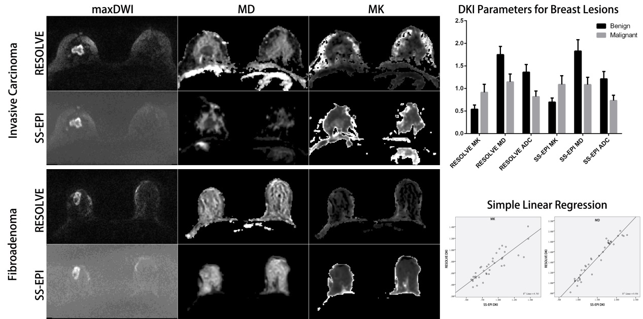
After approved by the institutional review board, 78 patients with 82 lesions (57 malignant and 25 benign) were scanned at 3T, using a 16-channel breast coil (Skyra, Siemens Healthcare). Two DKI sequences based on RESOLVE and SS-EPI were performed respectively. The diffusion-encoding gradients were applied in 3-orthogonal directions with four b-values (0, 50, 1000, 2000s/mm2). In RESOLVE sequence, 5 readout segments were applied. Mean Kurtosis (MK), mean diffusion (MD) and apparent diffusion coefficient (ADC) were calculated for each lesion on the two DKI data respectively. In addition, the image quality was assessed by a blinded reader, regarding the susceptibility artifact, image distortion, and anatomic details of breast lesions (margins and internal characteristics). The difference and correlation of both quantitative and qualitative parameters between RESOLVE and SS-EPI data were compared and statistically determined.Results:
In benign lesions, mean MK, MD (X 10−3mm2/s) and ADC (X 10−3mm2/s) were 0.540±0.094, 1.748±0.182 and 1.360±0.169 for RESOLVE data; 0.698±0.090, 1.828±0.251 and 1.213±0.164 for SS-EPI data. In malignant lesions, mean MK, MD and ADC were 0.914±0.177, 1.146±0.174 and 0.819±0.124 for RESOLVE DKI; 1.089±0.193, 1.086±0.160 and 0.732±0.117 for SS-EPI DKI. The differences were statistically significant for each corresponding parameters between two patient groups (P<0.001). The Pearson’s correlation showed a perfect linear relationship for each parameter between RESOLVE and SS-EPI (r = 0.874, 0.968 and 0.955 for MK, MD and ADC respectively; P<0.01 for all). In blinded read, RESOLVE DKI has significantly less susceptibility artifact/image distortion, and allowed for better evaluation of anatomic characteristics of breast lesions as compared with SS-EPI DKI, with very good agreement between two readers (Kappa = 0.85 – 0.94; P<0.01).Conclusions:
Applying RESOLVE technique can significantly improve the visualization of anatomic characteristics of breast lesions with less image distortion and higher image resolution, and its quantitative parameters are consistent with those from SS-EPI. Thus, RESOLVE DKI would be a useful diagnostic tool for breast diseases.Acknowledgements
I would like to show my deepest gratitude to Dr. Xu Yan, MR collaboration from Siemens Healthcare, for his valuable technical support and manuscript editing.References
1. Sun K, Chen X, Chai W, Fei X, Fu C, Yan X, Zhan Y, Chen K, Shen K, Yan F. Breast Cancer: Diffusion Kurtosis MR Imaging-Diagnostic Accuracy and Correlation with Clinical-Pathologic Factors. Radiology. 2015 Oct;277(1):46-55.
2. Wu D, Li G, Zhang J, Chang S, Hu J, Dai Y. Characterization of breast tumors using diffusion kurtosis imaging (DKI). PLoS One. 2014 Nov 18;9(11):e113240.
3. Nogueira L, Brandão S, Matos E, Nunes RG, Loureiro J, Ramos I, Ferreira HA. Application of the diffusion kurtosis model for the study of breast lesions. Eur Radiol. 2014 Jun;24(6):1197-203.
4. Porter DA, Heidemann RM. High resolution diffusion-weighted imaging using readout-segmented echo-planar imaging, parallel imaging and a two-dimensional navigator-based reacquisition. Magn Reson Med. 2009 Aug;62(2):468-75.
Figures