0934
Spiral-out and -in Double Echoes (SOIDE) Magnetic Resonance Fingerprinting with Improved T2 Mapping1Zhejiang University, Hangzhou, People's Republic of China, 2Massachusetts General Hospital
Synopsis
Magnetic resonance fingerprinting (MRF) provides fast and simultaneous mapping of parameters including T1 and T2. However, T2 mapping in MRF has been less robust and accurate than T1 in its current form. In this work, we propose a new sequence with spiral-out and -in double echoes (SOIDE) for MRF in each TR block which provide double echo signal with better T2 contrast. The proposed SOIDE-MRF shows improved performance over FISP-MRF, as demonstrated in Monte Carlo simulation on a numerical brain phantom (at Tacq=6s/slice, NRMSE of T2 reduced to 9.41% in SOIDE-MRF, and FISP-MRF ~11% at Tacq=12s/slice) and in vivo brain (less biased and more stable T2).
Purpose
MR fingerprinting1 is a novel technique which encodes multiple parameters in both spatial and temporal domain and then decodes then simultaneously with image reconstruction and dictionary matching. It has been implemented with trueFISP or FISP sequences to simultaneously achieve T1 and T2 mapping1,2. However, accurate parameterization normally requires long acquisition, especially for T2. To improve accuracy of T2 mapping and efficiency of MRF in general, a spiral-out and spiral-in double echoes (SOIDE) MRF is proposed and tested in both simulation and in vivo experiments.Methods
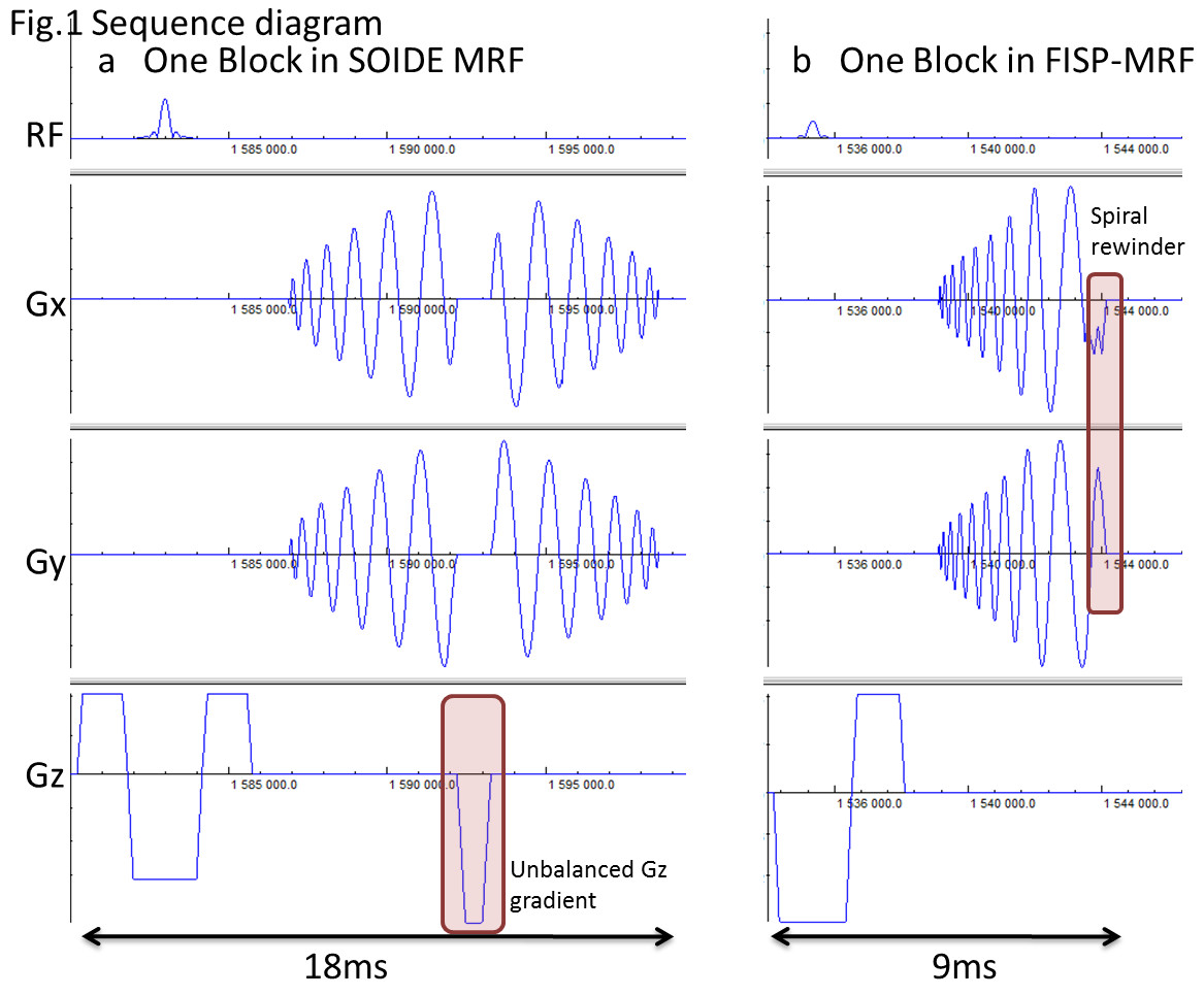
As shown in Fig1, adding upon the conventional FISP-based MRF, which has a single echo (spiral-out, short TE), the proposed SOIDE-MRF introduces a second echo (spiral-in, long TE). As such, it can generate an FID-like signal and also an echo-like signal in each TR, and provide better T2 information. In implementation, the second spiral trajectory in each TR is set to be the same as the first one with reversed order thus no dummy time is needed for the rewinder. Between the two spiral readouts, a Gz spoiler gradient is inserted as the unbalanced gradient. In the same manner as in FISP-MRF, the blocks vary in FAs and TRs to create temporal incoherence. The two echo signals from each block are reconstructed separately with a sliding window scheme3 and NUFFT reconstruction4 and then matched to the dictionary which is accurately modeled with extended phase graph (EPG) theory5. We used the same FA and TR time series for both SOIDE-MRF and FISP-MRF, and the same 36x undersampled spiral trajectory with a FOV of 22cm and 1.22mm voxel size. For SOIDE-MRF, 750 TRs were acquired with total acquisition time of 15s per slice. For comparison, 1500 TRs for FISP-MRF were acquired with the same 15s acquisition time.
To investigate the feasibility of the SOIDE-MRF, Monte Carlo simulation was conducted on a numerical brain phantom which includes the acquisition, sampling and reconstruction processes. Gaussian noise was added such that SNR=20 (for white matter). Simulations were repeated 50 times for statistical analysis which include pixel-wise and overall mean error and standard deviation calculation and also NRMSE. Experiments were also conducted on in vivo human brain with the same parameters at 3T(Siemens Prisma) using 20 channel receive coil.
Results
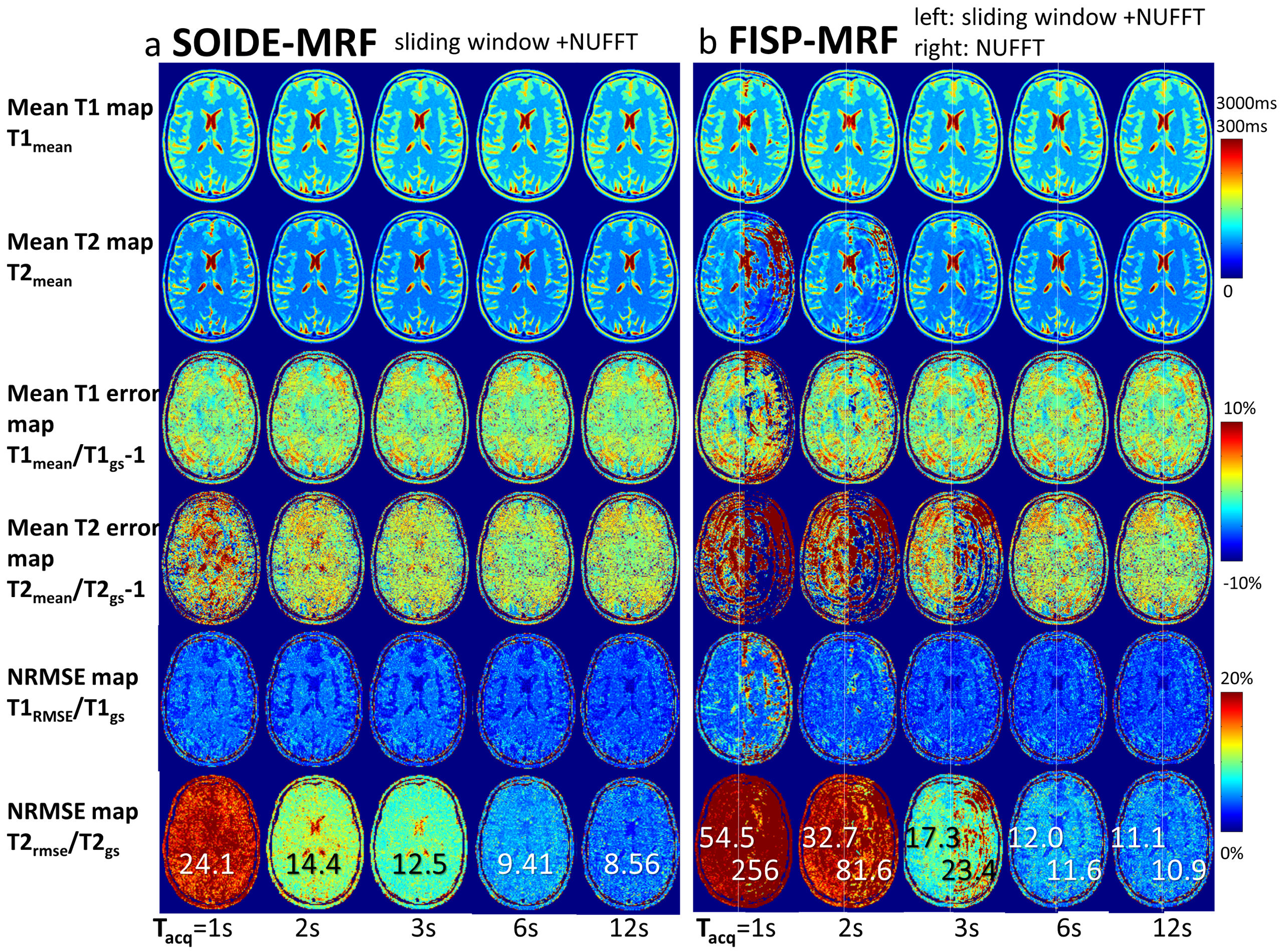
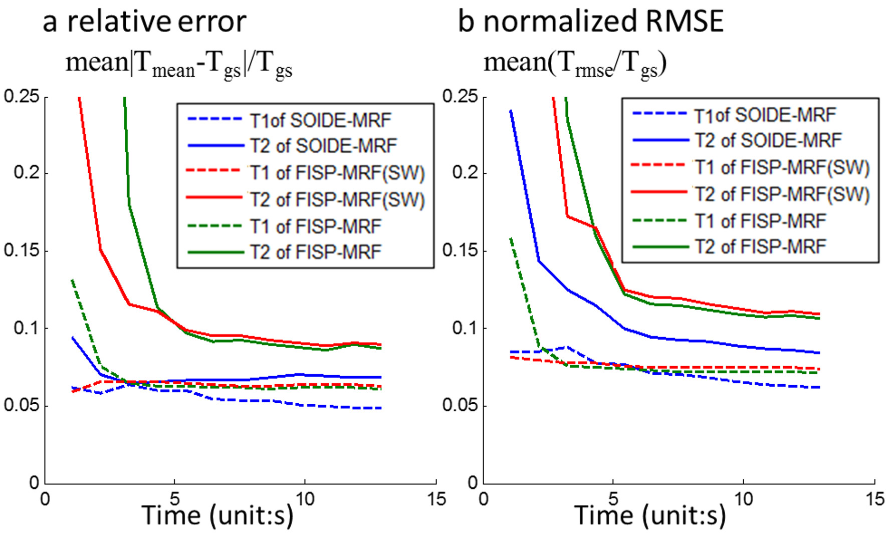
Fig 2 shows simulation results for both SOIDE-MRF and conventional FISP-MRF. For each case, the mean maps, mean error maps and normalized RMSE maps are shown for both T1 and T2 at different acquisition time, and the overall NRMSE of T2 is labeled in each panel. Fig.3. shows the overall relative error and normalized RMSE result versus acquisition time for different simulation cases.
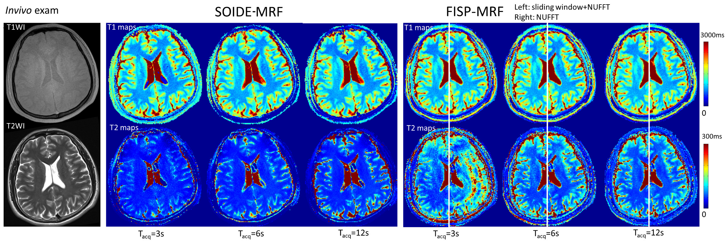
In vivo results are shown in Fig.4, which includes T1 and T2 maps for different acquisition durations, with conventional T1w and T2w images for comparison.
Discussion
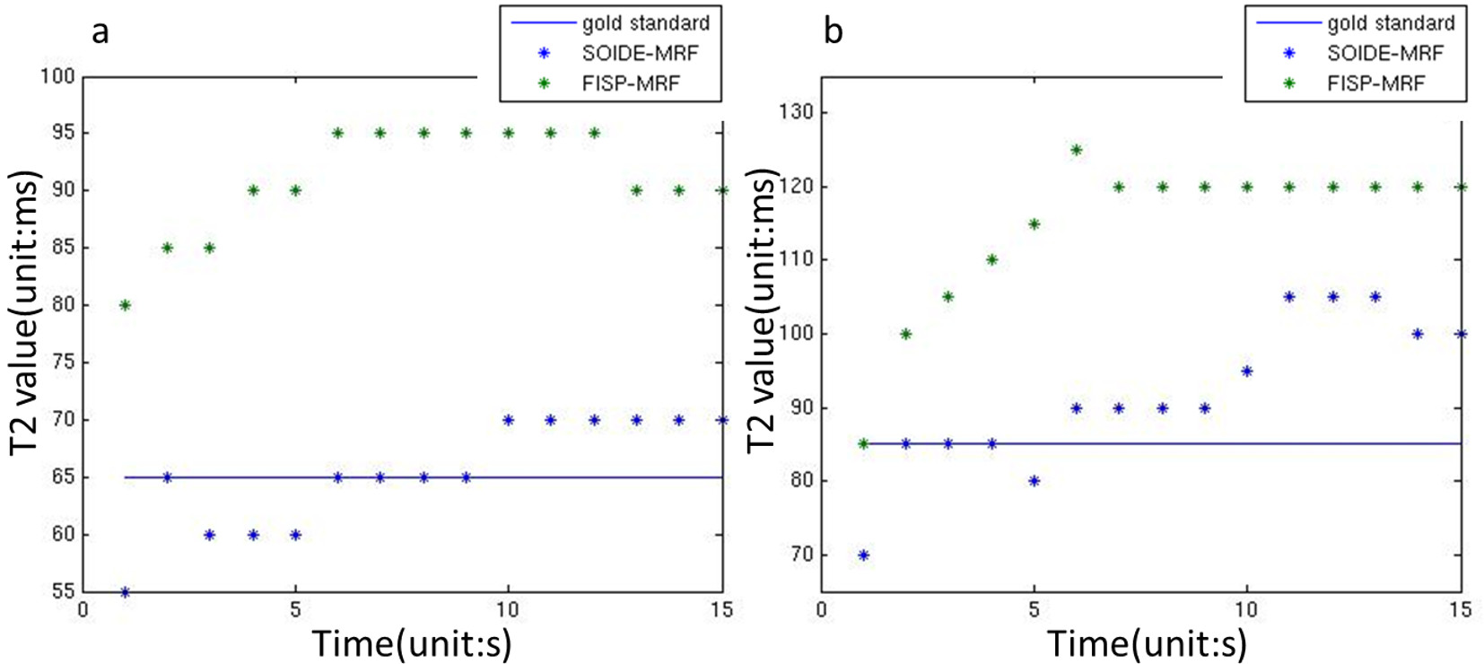
SOIDE-MRF shows significant improvements compared to conventional FISP-MRF especially for T2 quantification. Specifically, in simulation, for acquisition time larger than 2s/slice, SOIDE-MRF has an overall 7% mean error in T2 while for FISP-MRF the error is >9% for tacq<4s/slice. The overall NRMSE of T2 in SOIDE-MRF decreases >2%(at tacq>6s/slice) and >15%(at tacq<3s/slice) at all the acquisition time. This suggests the accuracy for T2 characterization is improved with SOIDE MRF. In vivo T2 mapping from SOIDE-MRF shows a closer value to that found in literature (at 3T, T2WM=56+4ms, T2GM=71+10ms)6 compared with that from FISP-MRF, in which the T2 over-estimation is likely a result of slice profile imperfection7. The decrease in SOIDE-MRF T2 indicates improved robustness to slice profile imperfection and good consistency; a claim that is supported by our simulation results in Fig.5.Conclusions
We have demonstrated that SOIDE-MRF provides accurate T1 and T2 maps with the proposed novel spiral-out and spiral-in strategy, which significantly outperform the current state-of-the-art FISP-MRF acquisition. Moreover, a Monte-Carlo simulation pipeline was developed and used in validating the MRF performance. Such simulation tool should prove useful for sequence and reconstruction design/optimization.Acknowledgements
This work is supported in part by the National Natural Science Foundation of China(No: 61427807,61271083, 61525106), by National Key Research and Development Program of China (No: 2016YFC1300302), and by Zhejiang Province Science and Technology Projects(No: 2015C33061).References
1. Ma D, Gulani V, Seiberlich N, et al. Magnetic resonance fingerprinting. Nature. 2013;495(7440):187-192. doi:10.1038/nature11971.
2. Jiang Y, Ma D, Seiberlich N, Gulani V, Griswold MA. MR fingerprinting using fast imaging with steady state precession (FISP) with spiral readout. Magn Reson Med. 2015;74(6):1621-1631. doi:10.1002/mrm.25559.
3. Cao X, Liao C, Wang Z, et al. Robust Sliding-Window Reconstruction for Accelerating the Acquisition of MR Fingerprinting. Magn Reson Med. 2016. doi:10.1002/mrm.26521(early view).
4. Fessler JA. On NUFFT-based gridding for non-Cartesian MRI. J Magn Reson. 2007;188(2):191-195. doi:10.1016/j.jmr.2007.06.012.
5. Weigel M. Extended phase graphs: Dephasing, RF pulses, and echoes - pure and simple. Magn Reson Med. 2014;00. doi:10.1002/jmri.24619.
6. Gelman N, Gorell JM, Barker PB, et al. MR Imaging of Human Brain at 3 . 0 T?: Preliminary Report on Transverse Relaxation Rates and Relation to Estimated Iron Content 1. Neuroradiology. 1999;210(3):759-767.
7. Ma D, Pahwa S, Gulani V, Griswold MA. Slice Profile Correction in 2D Magnetic Resonance Fingerprinting (MRF). In: Proc Intl Soc Mag Reson Med. ; 2016:3302.
Figures