0908
Treatment response of hepatic and renal stiffness in patients with chronic HCV infection monitored by multifrequency MR elastography1Department of Radiology, Charité - Universitätsmedizin Berlin, Berlin, Germany, 2Department of Medical Informatics, Charité - Universitätsmedizin Berlin, Berlin, Germany
Synopsis
Multifrequency abdominal MRE was applied to monitor stiffness of the liver and kidney in renal transplant recipients with chronic HCV infection after antiviral therapy. Within a 6-months period after medication, hepatic stiffness was significantly reduced while renal stiffness in kidney transplants was not altered. Wave speed obtained by high-resolution multifrequency MRE can serve as a quantitative imaging maker to assess the treatment response and monitor abdominal organ stiffness longitudinally.
Purpose
To noninvasively monitor stiffness of the liver and kidney in renal transplant recipients with chronic HCV infection after antiviral therapy using multifrequency MR elastography (MRE).Methods
In this prospective study, eight kidney transplant recipients with chronic HCV infection (age 52±14 years, 5 females) were treated with direct-acting antivirals (Daclatasvir/Sofosbuvir). They underwent MRE (1) examinations once before (day0) and twice at 3 months (3m) and 6 months (6m) after antiviral treatment. Multifrequency abdominal MRE (2) was performed on a 1.5-T scanner. Mechanical vibrations of 30, 40, 50, 60 and 70 Hz frequency were generated by pressure pads powered by medical compressed air. For each patient, two MRE experiments were conducted in axial and coronal slice orientations in order to cover the liver and the transplant kidney, respectively. Axial slice parameters were similar as detailed in (2): 4 vibration frequencies of 30 to 60 Hz, 11 consecutive slices of 2.7×2.7×5 mm3 resolution. The coronal protocol was the same as used in (3): 4 vibration frequencies of 40 to 70 Hz with, 11 contiguous slices of 2.5×2.5×2.5 mm3 resolution, 2 averages to improve SNR. Both coronal and axial scans were acquired under free breathing in a total acquisition time of 2 and 4 minutes, respectively. For each drive frequency, full 3D wave fields were recorded at eight time points of the vibration period. Data post-processing was based on the tomoelastography pipeline detailed in (4) which yields stiffness maps in terms of wave speed (in m/s). HCV load was assessed by real-time PCR over 6 months period.Results
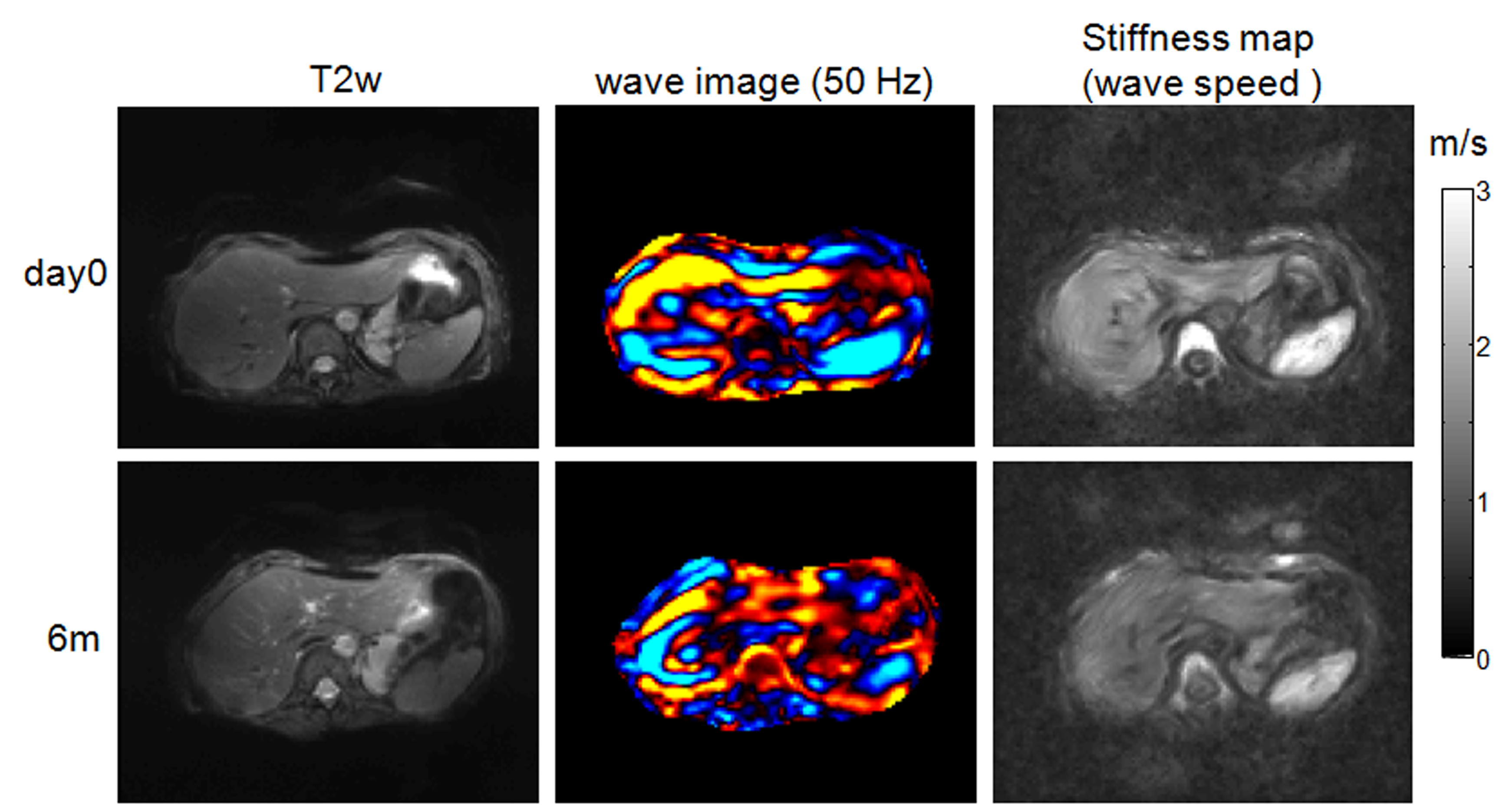
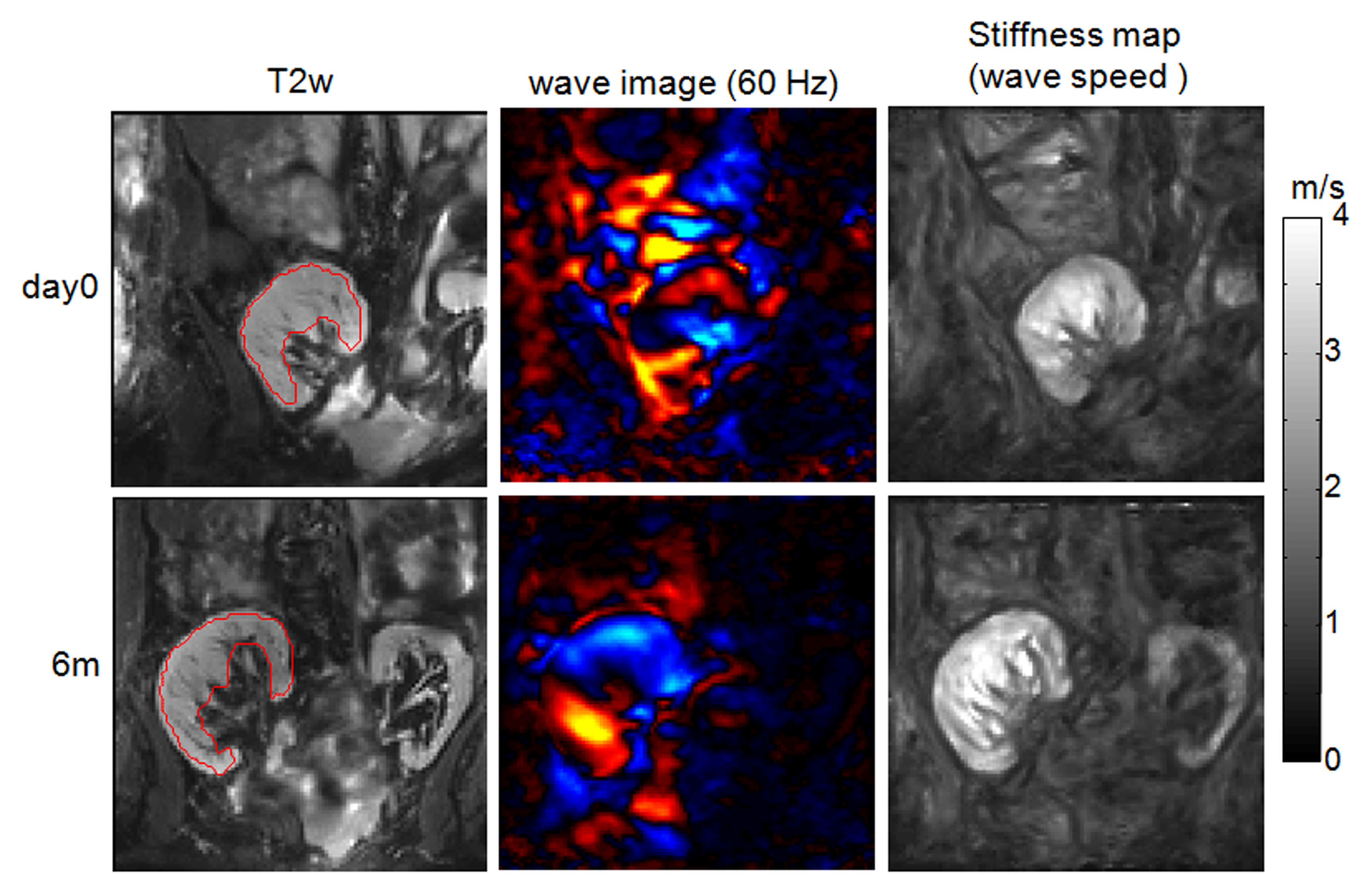
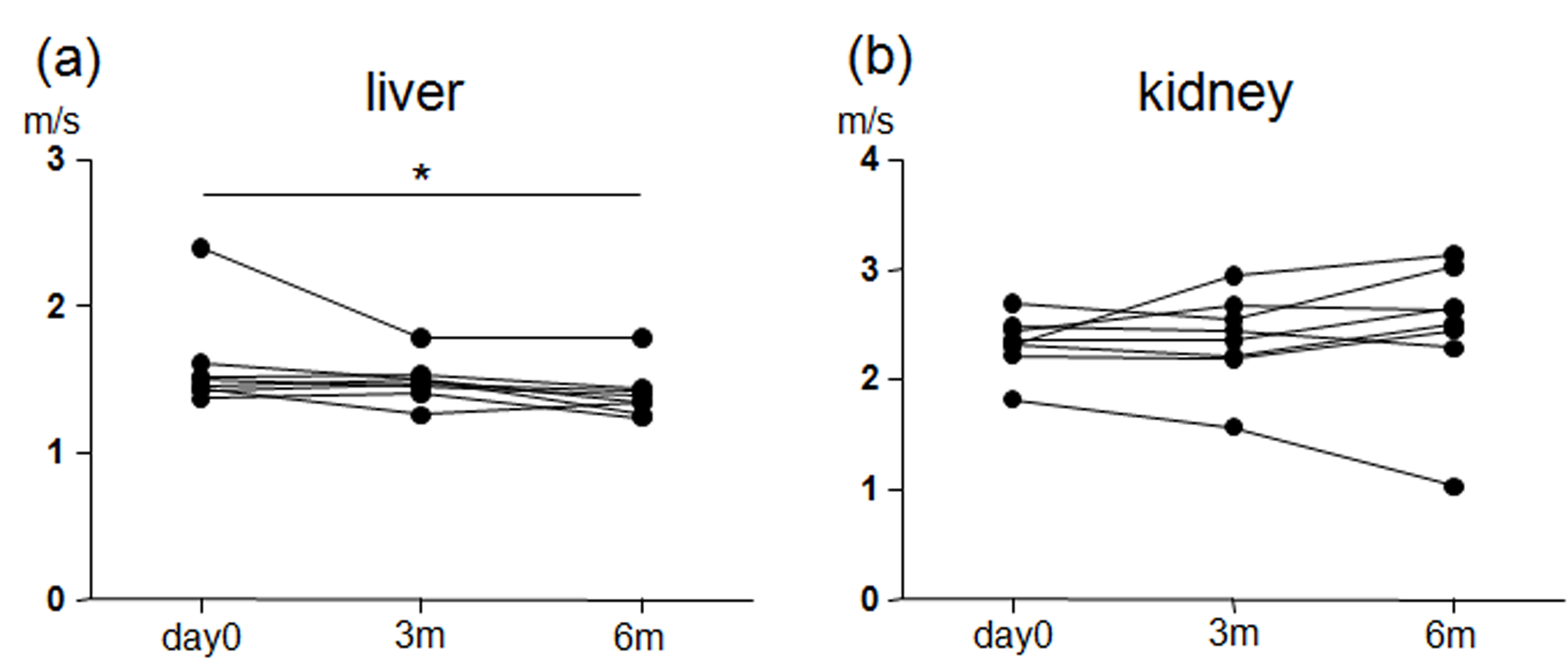
MRE magnitude images (T2w), in-plane shear strain images (wave image) and the corresponding stiffness map (wave speed) are shown for liver (Fig.1) and the transplant kidney (Fig.2) in the same patient at day0 and 6 months. HCV load was reduced by treatment below detectable thresholds after 19 days (range 8–29 days). MRE showed a significant decrease in liver stiffness after 6 months (day0, 1.59±0.33 m/s; 3m, 1.49±0.15 m/s; 6m, 1.40±0.17 m/s; P<0.05; repeated-measure ANOVA). No significant differences were observed in the transplant kidneys during the 6-months periods (day0, 2.33±0.25 m/s; 3m, 2.37±0.40 m/s; 6m, 2.47±0.64 m/s; P=0.36; repeated-measure ANOVA),. The stiffness of both liver and the transplant kidney at all three time points are plotted in Fig.3a and Fig.3b, respectively.Discussion
In this prospective longitudinal study, high-resolution multifrequency MRE was applied to monitor the stiffness of both the liver and renal transplants in patients with HCV after antiviral treatment. To our knowledge, this is the first MRE study to monitor the treatment response of liver and transplant kidney to effective antiviral medication. Previous studies addressed liver stiffness in patients with HCV by MRE for fibrosis staging (5, 6) and for the assessment of cirrhosis progression (7) but did not investigate therapy effects. Our data show that reduction of HVC load does not immediately alter hepatic stiffness. The observed long-term changes of hepatic stiffness suggest that remodeling of the liver occurs further downstream the pathophysiological cascade. In the context of kidney transplant, we found no significant changes in renal stiffness of the transplant kidney, which suggests that the drug metabolism of the combined Daclatasvir and Sofosbuvir didn't influence the mechanical property of the renal transplants in the course of 6 month. These preliminary findings might provide reference data for MRE in HVC therapy monitoring and other applications of clinical pharmacology. More patients and longer monitoring periods are planned for this study.Conclusion
Stiffness maps obtained by high-resolution multifrequency MRE can serve as a quantitative imaging maker to assess the treatment response and monitor abdominal organ stiffness longitudinally. Within a 6-months period after antiviral therapy, hepatic stiffness was significantly reduced while renal stiffness in kidney transplants was not altered.Acknowledgements
No acknowledgement found.References
1. Muthupillai R, Ehman RL. Magnetic resonance elastography. Nat Med. 1996;2(5):601-3.
2. Dittmann F, Tzschätzsch H, Hirsch S, et al. Tomoelastography of the abdomen: Tissue mechanical properties of the liver, spleen, kidney and pancreas from single MR elastography scans at different hydration states. Magn Reson Med. 2016;DOI 10.1002/mrm.26484.
3. Marticorena Garcia SR, Fischer T, Dürr M, et al. Multifrequency Magnetic Resonance Elastography for the Assessment of Renal Allograft Function. Invest Radiol. 2016;doi:10.1097/RLI.0000000000000271.
4. Tzschatzsch H, Guo J, Dittmann F, et al. Tomoelastography by multifrequency wave number recovery from time-harmonic propagating shear waves. Med Image Anal. 2016;30:1-10.
5. Ichikawa S, Motosugi U, Ichikawa T, et al. Magnetic resonance elastography for staging liver fibrosis in chronic hepatitis C. Magn Reson Med Sci. 2012;11(4):291-7.
6. Shire NJ, Yin M, Chen J, et al. Test-retest repeatability of MR elastography for noninvasive liver fibrosis assessment in hepatitis C. J Magn Reson Imaging. 2011;34(4):947-55.
7. Takamura T, Motosugi U, Ichikawa S, et al. Usefulness of MR elastography for detecting clinical progression of cirrhosis from child-pugh class A to B in patients with type C viral hepatitis. J Magn Reson Imaging. 2016;44(3):715-22.
Figures