0891
Differential diagnosis of Hepatic metabolites between non-alcoholic steatohepatitis and simple steatosis in humans and a murine model using a 1H MR spectroscopy study with long TE1Radiology, Imaging Science Research Center, Iksan, Korea, Republic of, 2Radiology, Wonkwang University School of Medicine, Iksan, Korea, Republic of
Synopsis
Liver biopsy is the gold standard for diagnosing non-alcoholic fatty liver disease(NAFLD) but with practical constraints. MR spectroscopy(MRS) allows in vivo assessment of hepatocellular metabolism and has shown potential for biochemical differentiation in diffuse liver disease. Recent 13C-MRS demonstrated that alanine and lactate levels in inflammatory liver injury were increased compared to those in normal liver, and these changes were positively correlated with liver enzyme levels. However, the clinical use of 13C-MRS technique is limited because it necessitates 13C-resonance specific hardwares and softwares for data acquisition. This study used long TE 1H-MRS to monitor changes of hepatocellular metabolites in NAFLD.
Purpose: Liver biopsy is the gold standard for diagnosing non-alcoholic fatty liver disease(NAFLD) but with practical constraints. Proton MR spectroscopy(1H-MRS) allows in vivo assessment of hepatocellular metabolism and has shown potential for biochemical differentiation in diffuse liver disease. This study was evaluated the usefulness of hepatic metabolic changes determined by 1H-MRS to differentiate non-alcoholic steatohepatitis(NASH) from simple steatosis in human and a murine model.
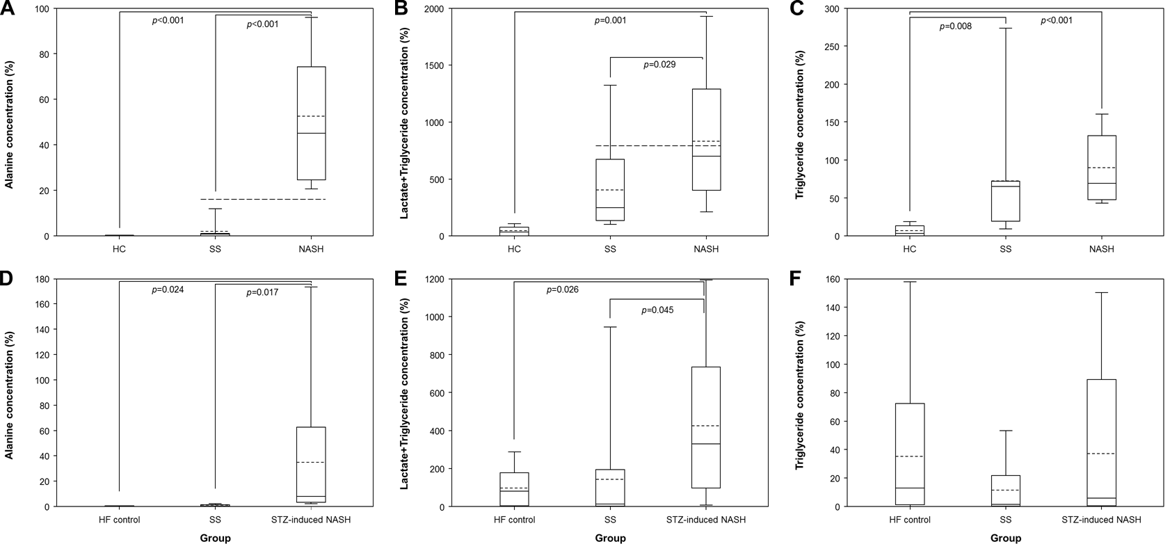
Methods: A total of thirty-two subjects─11 patients with NASH, 15 with simple steatosis, and six healthy controls─were studied. In order to test the reproducibility, thirty-six C57BL/6 mice, including 10 mice with streptozotocin-induced NASH, 15 with simple steatosis, and 11 high-fat diet controls, were studied. 1H-MRS measurements were performed on a localized voxel of the liver using a point-resolved spectroscopy(PRESS) sequence. Hepatic alanine(Ala), lactate+triglyceride (Lac+TG), and TG levels were compared between NASH, simple steatosis and control groups using ANOVA and Tukey’s post-hoc tests. Diagnostic accuracy was determined by calculating the area under the receiver operating characteristics(ROC) curve. The associations between metabolite levels and pathologic grades or NAFLD activity scores were assessed using Pearson’s correlation.
Results: NASH patients had higher levels of Ala (p<0.001), Lac+TG (p<0.001), and TG (p<0.05) than simple steatosis patients or controls. The area under the ROC curve to distinguish NASH from simple steatosis was 1.00(95% confident interval (CI) 1.00–1.00) for Ala and 0.782 (95% CI 0.61–0.96) for Lac+TG. Ala and Lac+TG levels correlated well with steatosis grade, lobular inflammation, and NAFLD activity scores. The metabolic changes in humans were reproducible in a mouse model of liver disease.
Conclusion: 1H-MRS is potentially useful for non-invasive diagnosis of NASH and simple steatosis by hepatic metabolite quantification.
Acknowledgements
This study was supported by a grant of the Korean Health Technology R&D Project, Ministry of Health & Welfare, Republic of Korea.References
1. Csak T, Ganz M, Pespisa J, Kodys K, Dolganiuc A, Szabo G. Fatty acid and endotoxin activate inflammasomes in mouse hepatocytes that release danger signals to stimulate immune cells. Hepatology. 2011; 54(1): 133-44.
2. Lee SS, Park SH. Radiologic evaluation of nonalcoholic fatty liver disease. World J Gastroenterol. 2014; 20(23): 7392-402.
3. Pineda N, Sharma P, Xu Q, Hu X, Vos M, Martin DR. Measurement of hepatic lipid: high-speed T2-corrected multiecho acquisition at 1H MR spectroscopy--a rapid and accurate technique. Radiology. 2009; 252(2): 568-76.
4. Josan S, Billingsley K, Orduna J, et al. Assessing inflammatory liver injury in an acute CCl4 model using dynamic 3D metabolic imaging of hyperpolarized [1-(13) C]pyruvate. NMR Biomed. 2015; 28(12): 1671-7.
Figures
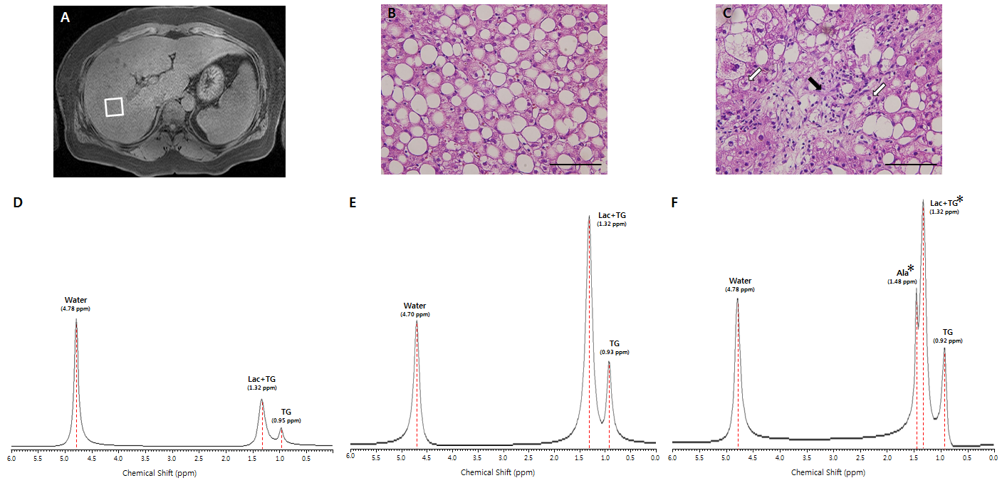
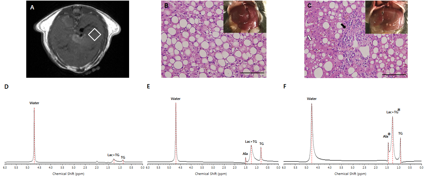
Figure 1. An axial plane anatomic image with spectroscopy voxel localized in the right lobe of liver from a patient (A) and photomicrographs showing the histopathological features of liver in patients with simple steatosis (B) and NASH (C) (H&E staining, ×400 magnification). Representative 1H MR spectra with 288 ms TE in a healthy control subject (D) and patients with simple steatosis (E) and NASH (F).