0846
A preliminary application of Porosity Index measured by UTE MRI sequence in the femoral neck1Radiology Department of Peking University Third H, Beijing, People's Republic of China, 2GE Healthcare, MR Research China, BeiJing, People's Republic of China
Synopsis
The current study aims to assess the feasibility of porosity index (PI) measurements derived from ultra-short echo time (UTE) MRI technology in the femoral neck and to further investigate its latent associations with age, gender, body mass index (BMI) and tibial PI. It was concluded that cortical PI measured by UTE MRI sequence can be applied in femoral neck and cannot be replaced by tibial measurement. Femoral neck and tibial PI were observed to correlate with age and BMI , which worth further study.
Purpose
The purpose of this study is to assess the feasibility of the measurements of porosity index (PI) measurements derived from ultra-short echo time MRI (UTE MRI) in the femoral neck and to further investigate its latent associations with age, gender, body mass index (BMI) and tibial PI.Introduction
Cortical bone accounts for 80% of human bones[1] and plays a significant role on defining cortical bone quality especially in bone fractures of OP patients. The recently developed ultrashort TE (UTE) MRI technology, with an echo time less than 0.5ms,is able to detect cortical bone signal and provide information of cortical bone microarchitecture. Rajapakse[2] lately developed porosity index (PI) as a surrogate of tibia cortical porosity by measuring the signal decay at only two TEs on a clinical 3T MR scanner. The current study applied this two-point UTE sequence in the femoral neck to investigate the relation of femoral neck PI and age, gender, BMI in healthy people. The potential association between femoral neck PI and tibial PI was further investigated to ensure the plausibility of tibial PI results on reflecting modifications in the femoral neck.Material and Methods
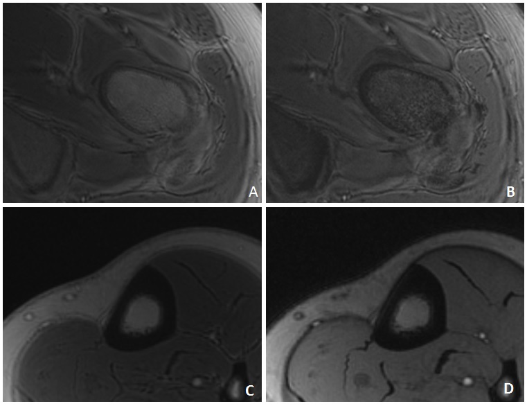
68 healthy volunteers (age 46±16 years, age range 21-76 years) were recruited while excluding those with fragile fracture histories, long-time limitation of normal physical activity and medical histories including diseases or treatments that may affect bone metabolism. All the volunteers underwent double-echo 3D UTE MRI examinations of hip and tibia on a 3.0T MR station (MR750w, GE Healthcare). The UTE sequence parameters were as follows: For tibia, TE1=0.1ms as the minimum TE on the scanner, TE2=4ms, TR=12ms, FOV=12cm×12cm, Bandwidth=62.5Hz, NEX=2, Flip angle=12°, slice-thickness=2.4mm, reconstruction matrix=256×256, and scanning time = 4.5mins. For Hip, only different parameters from tibia scan were displayed as TE2=4.6ms, TR=12.2ms, FOV=17cm×17cm, reconstruction matrix=384× 384, and scanning time = 6.5mins. Porosity Index of the cortical area was calculated as the ratio between TE2 and TE1 UTE image intensities, namely: porosity index (%) = (TE2 intensity/TE1 intensity) · 100%. The ratio images were post-processed using FuncTool on GE AW4.6 workstation out of which the slice with the thickest cortex was selected. The whole cortical bone region was analyzed for the tibia, while the inferior region of femoral neck cortex was analyzed as it was the thickest region.Results
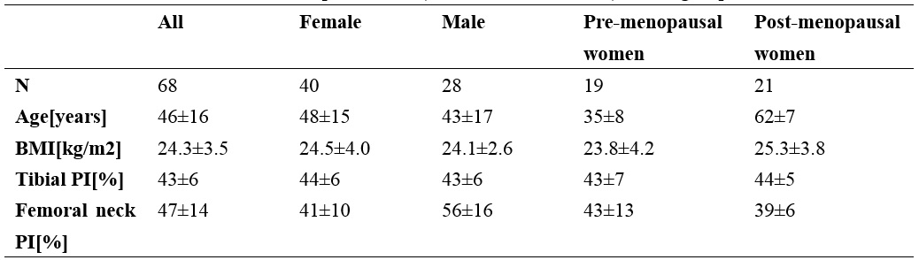
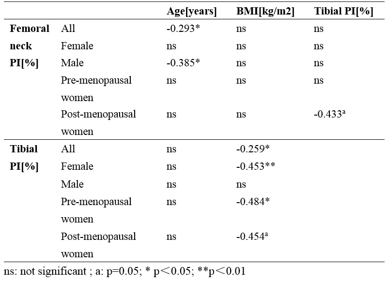
The femoral neck and tibial UTE images were successfully obtained from all the healthy volunteers (Figure 1). The characteristics of the parameters for different groups were shown in table 1.Tibial PI and femoral neck PI were not correlated over all age groups but displayed an association in premenopausal women(r=-0.433, p=0.050). Femoral neck PI demonstrated a negative correlation with age in men (r=-0.385, p<0.05) but an absence of correlation with BMI in all groups. Tibial PI was significantly correlated with BMI in female(r=-0.453,p<0.01),without any correlation with age in all groups. However, tibial PI displayed a positive correlation with age in postmenopausal women(r=0.429, p=0.067). In addition, male femoral PI was significantly higher than that of the female (p<0.001), but no difference between pre- and post-menopausal women was observed. The results were summarized in table 2.Discussion and Conclusion
To our best knowledge, this is a preliminary study of applying PI measured by UTE MRI in femoral neck. It is concluded that PI of the femoral neck can be achieved by using a clinical 3.0T MR workstation with a 6.5 mins UTE sequence. Tibial PI was not correlated with the femoral neck PI in most of the groups, indicating the necessity of direct assessment of the femoral neck cortex while estimating bone quality.The inter-gender difference in the femoral neck PI is consistent with the existing literatures. The negative correlation observed between tibial PI and BMI can be attributed to the mechanical stimulation; whereas the observed decrease of femoral neck PI with age was in contrary to most of the previous literatures on cortical porosity. However, significant individual variations in cortical bone porosity were observed by Stein and his colleagues[3], with low porosity in some of the elderly subjects and high porosity in some of the young adults.Thus further study is still needed.Acknowledgements
No acknowledgement found.References
[1]
Du, J, G. M. Bydder. Qualitative and quantitative ultrashort-TE MRI of cortical
bone. NMR Biomed 26(5): 489-506.
[2]
Rajapakse, C. S,Bashoor-Zadeh, M,Li, C, et al. Volumetric Cortical Bone
Porosity Assessment with MR Imaging: Validation and Clinical Feasibility.
Radiology 276(2): 526-535.
[3] Stein MS, Feik SA, Thomas CD ,et al.An
automated analysis of intracortical porosity in human femoral bone across age.
J Bone Miner Res 14(4):624–632
Figures