0780
Motion corrected high resolution time-of-flight angiography at 7T using Segmented FatNavs1EPFL, Lausanne, Switzerland, 2School of Engineering, Cardiff, United Kingdom
Synopsis
We propose to insert Segmented FatNavs into a time-of-flight sequence, and use it as a multi-purpose module. It allows fat suppression, magnetization-transfer suppression of the tissue signal, and retrospective motion correction. Image quality was always enhanced after correction, even in cases of small motion, thereby making high-resolution brain angiography more reliable.
Introduction
Very high spatial resolution MRI scans are challenging due to many limitations, including lower SNR, increased sensitivity to motion and longer scan-times. Fat navigators (FatNavs) are a highly-accelerated fat selective 3D GRE acquisition, and they have shown excellent suitability for motion tracking and retrospective correction1,2.
Time-of-flight (TOF) sequence data is usually visualized using a maximum intensity projection (MIP), and vessels close to one another along the projection can become difficult to distinguish when blurred by even small motion.
We propose using a segmented version of the FatNavs (SegFatNavs)3 with a TOF sequence to allow retrospective motion correction. The fat-selective binomial pulse has the additional advantage of acting as a fat-suppression pulse for the TOF as well as a magnetization transfer (MT) pulse, which has been shown to help suppress background tissue in TOF imaging4. The fat-suppression and MT effects are easily tuneable by adjusting the excitation flip angle of the SegFatNavs, as sufficient SNR is available even when far from the Ernst angle.
Methods
We used a 7T head-only MR scanner (Siemens Healthcare, Germany) and a 32-channel RF coil (Nova Medical Inc.). Three healthy volunteers were asked to stay still for the duration of the scan. We acquired two axial high-resolution datasets, with an isotropic voxel size of 0.4mm and 0.25mm respectively. The imaging slabs were 8.3cm and 4cm respectively (chosen to cover the Circle of Willis), with the 0.25mm protocol limited by memory restrictions of the current sequence implementation.
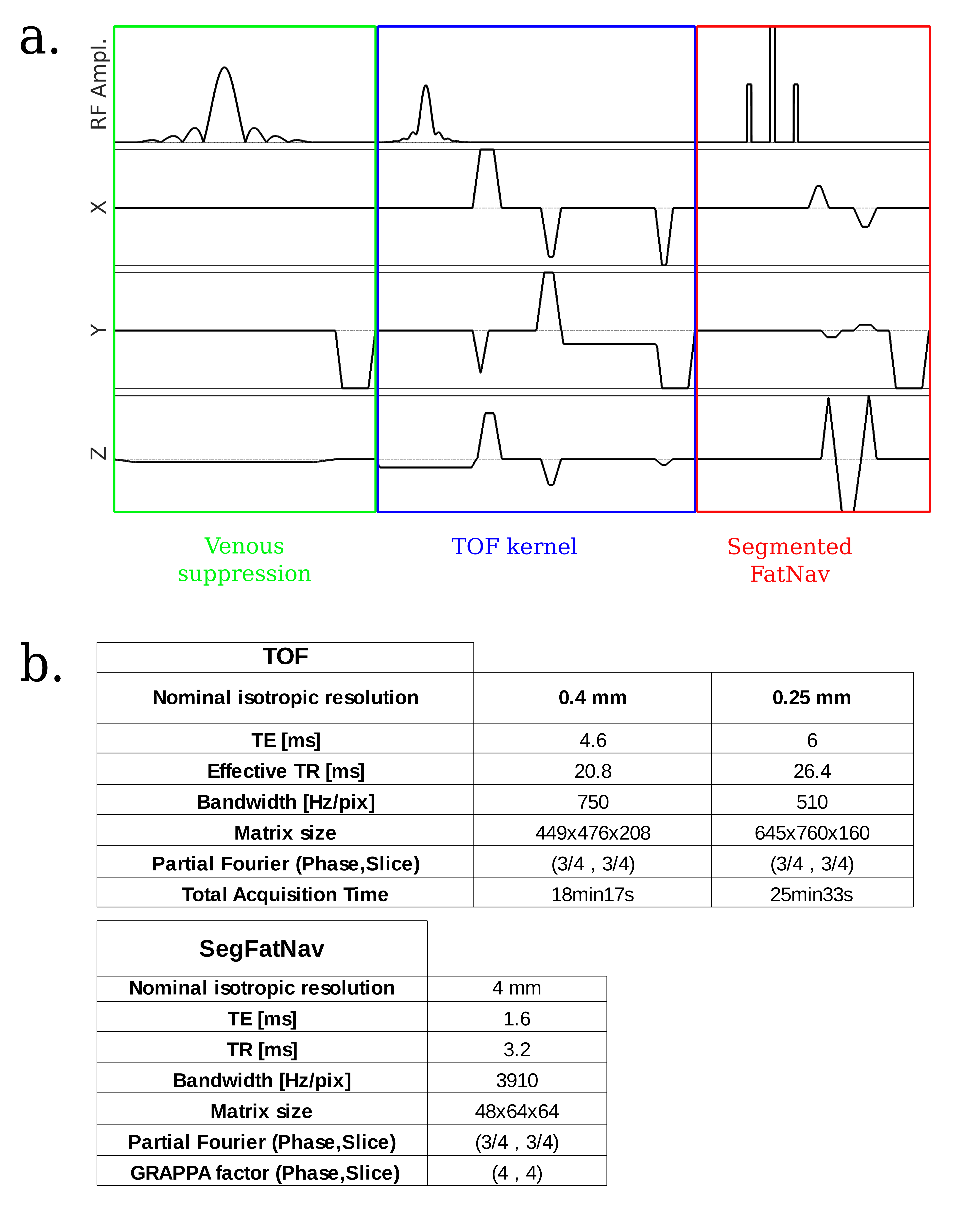
The custom-made sequence kernel (Figure 1a) included a venous saturation pulse, a TONE5 pulse followed by a fully flow-compensated GRE acquisition, and the SegFatNavs module. The nominal TONE flip angle was 20° and the slope factor was 50% in the foot-head direction. The SegFatNavs flip angle was chosen as high as possible while still respecting standard SAR constraints. Other parameters values can be found on Figure 1b. GRAPPA reference lines for the SegFatNavs were acquired in a 2.3s pre-scan for each dataset. One full navigator volume was acquired every 2.25s / 2.85s for the 0.4mm / 0.25mm scan, respectively. SegFatNavs were retrospectively co-registered using SPM to obtain the time-courses of the rigid body motion parameters, and retrospective motion correction of the time-of-flight sequence was performed coil-wise using a NUFFT6 adjoint operator approach.
Finally, MIPs were performed on sum-of-squares images, and brain masks (if used) were always obtained from the motion corrected reconstruction for fairer comparisons.
Results and discussion
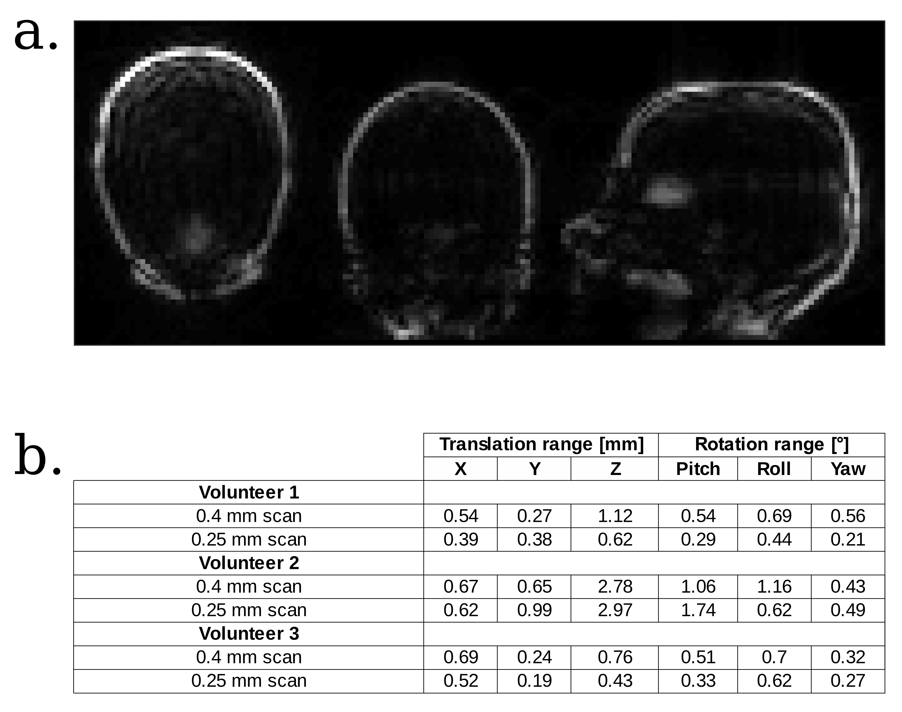
An example of a SegFatNav volume is shown in Figure 2a, and the range of the estimated motion for all scans (calculated independently for each motion parameter) is presented on Figure 2b. Volunteers 1 and 3 remained very still, whereas Volunteer 2 exhibited some mild motion.
The uncorrected and corrected images of a 0.4mm scan are compared on Figure 3, where the sagittal MIP was performed after brain extraction. The vessels sharpness is greatly increased in the corrected reconstruction.
Figure 4 shows the 0.25mm scans results: the top image (volunteer 1) demonstrates excellent venous and fat suppressions, while the bottom movie (volunteer 3) shows that even with small motion (translations < 0.53 mm, rotations < 0.63°), vessels have higher signal and better contrast after correction, and hence are more easily visualized.
Conclusion
The inclusion of SegFatNavs into the TOF sequence enables greatly improved robustness of image quality in the face of subtle subject motion. Future work will aim to quantify under which conditions the magnetization transfer effect induced by the fat excitation pulse further enhances vessels to static tissues contrast. Finally, due to the modularity of the SegFatNavs approach, the sequence can also be extended to a multi-slab version.Acknowledgements
This work was supported by CIBM of the UNIL, UNIGE, HUG, CHUV, EPFL and the Leenaards and Jeantet Foundations, as well as SNSF project number 205321_153564.References
1. Gallichan D, Marques JP, Gruetter R. Retrospective correction of involuntary microscopic head movement using highly accelerated fat image navigators (3D FatNavs) at 7T. Magnetic Resonance in Medicine. 2015;0:1–10.
2. Skare S, Hartwig A, Mårtensson M, Avventi E, Engström M. Properties of a 2D fat navigator for prospective image domain correction of nodding motion in brain MRI. Magnetic resonance in medicine. 2015;73(3):1110–9.
3. Gretsch F, Kober T, Waszak M, Marques JP, Gallichan D. High temporal resolution retrospective motion and B0 correction using FIDNavs and segmented FatNavs at 7T. In: Proc. Intl. Soc. Mag. Reson. Med. 24. 2016. p. 4251.
4. Pike GB, Hu BS, Glover GH, Enzmann DR. Magnetization transfer time-of-flight magnetic resonance angiography. Magnetic resonance in medicine?: official journal of the Society of Magnetic Resonance in Medicine / Society of Magnetic Resonance in Medicine. 1992;25(2):372–379.
5. Atkinson D, Brant-Zawadzki M, Gillan G, Purdy D, Laub G. Improved MR angiography: magnetization transfer suppression with variable flip angle excitation and increased resolution. Radiology. 1994;190(3):890–4.
6. Fessler JA, Sutton BP. Nonuniform Fast {Fourier} Transforms Using Min-Max Interpolation. IEEE \Trans\ Signal Processing. 2003;51(2):560–574.
Figures