0779
Prospective motion correction on diffusion weighted imaging: improving data quality with four radio frequency and gradient pulses updates.1University of Hawaii, Honolulu, HI, United States, 2Department of Radiology, University Medical Center Freiburg, Freiburg, Germany, 3Department of Medicine, John A. Burns School of Medicine, University of Hawaii, Honolulu, HI, United States
Synopsis
Prospective Motion Correction (PMC) using fast camera-based tracking systems can dramatically increase DWI data quality and may have an important application in populations where movement is hard to prevent. Here we present images acquired with and without intra-sequence PMC from two subjects realizing fast head rotations up to 30°/s. Our results show that the DWI motion sensitivity can be reduced five-fold by applying 3 intra-sequence PMC updates (prior to each of the two refocusing RF pulse and prior to readout).
Purpose
The quality of brain Diffusion Weighted Imaging (DWI) is affected by head motion. Motion effects are commonly thought of in terms of slice misalignments, which can be eliminated using prospective motion correction (PMC). However, rotations during diffusion-sensitizing gradients can induce spurious gradient moments, and therefore shifts in k-space1. Thus, even when slices are aligned properly, the MR signal can be lost (signal dropout) if the spurious gradient moment is sufficient to shift the echo center out of k-space. Fast PMC with optical tracking (≥50 frames/second) can resolve these issues, by correcting spurious gradient moments. Two different approaches have been demonstrated: 1) apply quasi-continuous PMC updates by segmenting and updating diffusion-gradient waveforms every 2ms1, and 2) update slice positions, calculate spurious gradient moments based on multiple tracking data and gradient timings, and apply gradient blips prior to data acquisition to reverse spurious gradient moments2. However, the implementation of these approaches can be challenging and may not be feasible on all platforms. Here, we examine whether signal dropouts during DWI can be eliminated by applying only three intra-sequence PMC updatesMethods
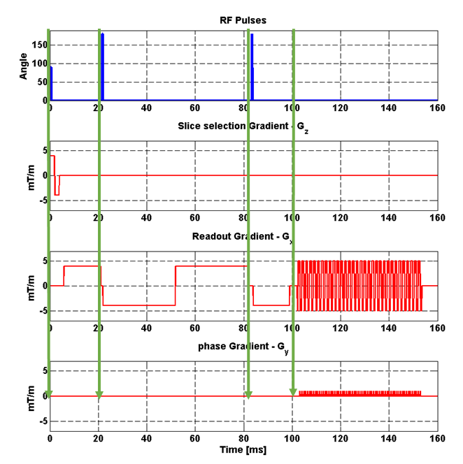
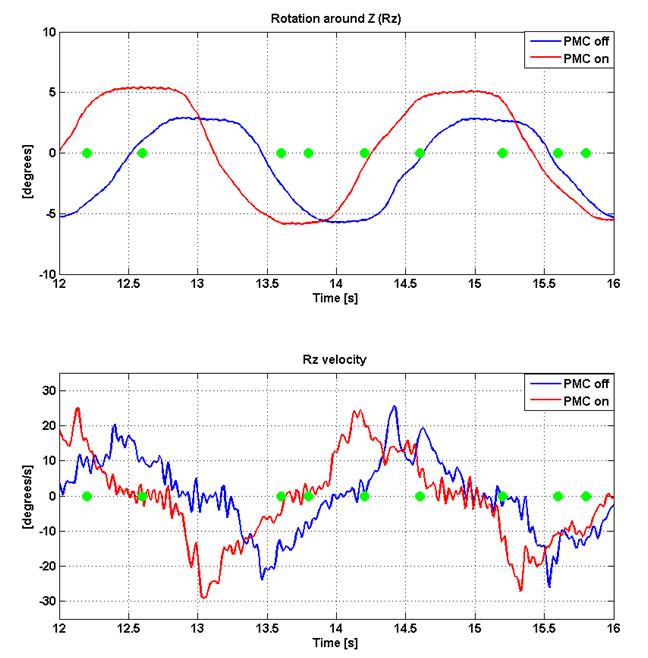
Studies were performed on a 3T Trio (Siemens, Germany) with single-shot dual-echo EPI [20 slices (3x3x3mm3), matrix 64x64, TE/TR=87/4000ms, bandwidth=1502Hz/pixel, b=500s/mm2 in readout direction]. One measurement was performed with phase 6/8 partial-Fourier (Subject 1), and one without Partial-Fourier (Subject 2). An MR-compatible tracking system (Metria, USA) was used to track head motion at 80 frames/s, using a Moiré marker3 attached to subjects’ forehead. The high frame rate made it possible to apply 3 intra-sequence PMC updates (prior to each refocusing pulse and prior to readout, Fig.1) and estimate the head velocity for each excitation. Tracking data were synchronized with DWI timing via unique packet IDs. In each of 2 male subjects, four DWI data sets were acquired with the following conditions: subject keeping still without PMC (PMC-OFF), left-right head movement with PMC-OFF, and repeated both with PMC-ON. Five volumes were acquired per condition. Subjects were trained to execute the side-to-side motion before the scan to reproduce amplitude and velocity across scans.Results
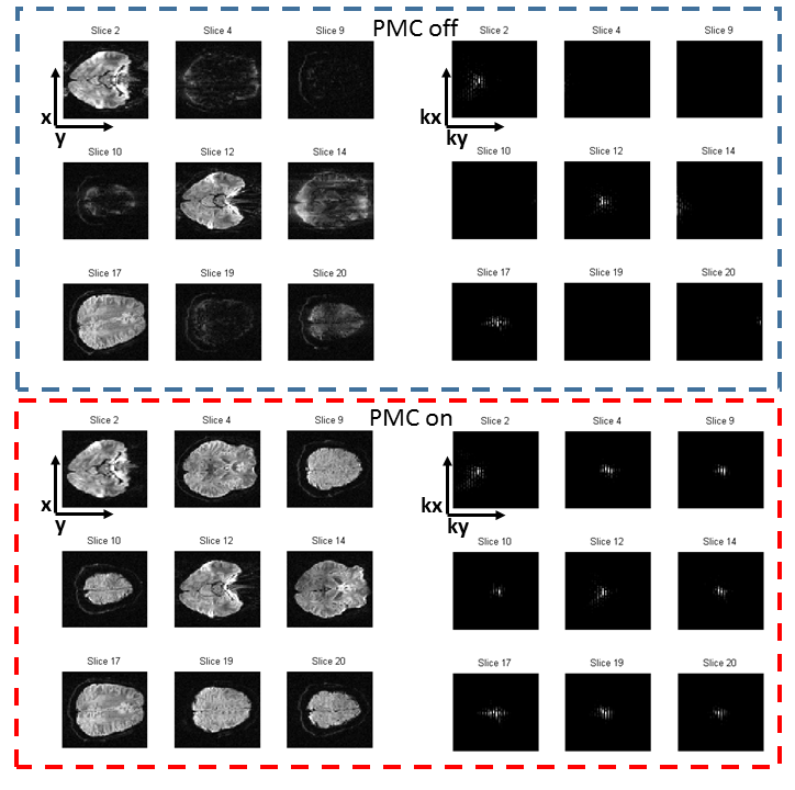
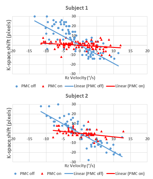
Echo shifts are much more pronounced when PMC is OFF compared to ON (Fig. 2, right column top versus bottom). For example, at Vz~10°/s (green dots, Fig. 3) for PMC-OFF (slice 19, Fig. 2), the signal was pushed to the edge of k-space, leading to partial signal loss. Conversely, the echo center was barely shifted for PMC-ON acquisitions at similar velocities (slice 20). For higher velocities (~15°/s), there is complete signal loss with PMC-OFF (slice 9), whereas no significant echo shift or artifacts are visible for PMC-ON (slice 12; rotational velocity ~20°/s). Likewise, the regression slope of k-space shifts versus z-rotation velocity is decreased for PMC-ON acquisitions in both subjects (PMC-OFF and PMC-ON average slope -1.92 and -0.30[pixels*s/deg], Fig. 4). For full-Fourier acquisitions with PMC-OFF (Subject 2), the echo center was shifted to the edge of k-space at approximately 20°/s Vz. Conversely, PMC-ON acquisitions may tolerate velocities over 5 times higher (>100°/s). Subject 1 also exemplifies that images acquired with partial Fourier methods are more strongly affected by head motion (negative k-space showing signal loss for Vz ≥8°/s).Discussion
We demonstrate that a simpler approach for applying intra-sequence PMC updates (n=3 updates only) can be effective for attenuating the motion-sensitivity of DWI. Real time corrections are particularly useful for partial-Fourier acquisitions, which are yet more sensitive to motion due to the reduced size of k-space. Furthermore, the sensitivity of DWI to movements is generally proportional to b-values applied; therefore, the five-fold reduction in motion-sensitivity afforded by our method might be particularly useful for high b-value acquisitions1. Finally, our approach may be improved further by adding an extra PMC update between the two refocusing pulses.Conclusion
We demonstrate a simple real-time approach of applying PMC sequence updates, which decreases DWI motion-sensitivity about 5-fold. PMC updates are particularly important for acquisitions using partial-Fourier and those with high b-values.Acknowledgements
No acknowledgement found.References
1-Herbst M, Maclaren J, Weigel M, et al. Prospective motion correction with continuous gradient updates in diffusion weighted imaging. Magn Reson med. 2012;67,326-338.
2-Gumus K, Keating B, Poser BA, et al. Prevention of motio-induced signal loss in diffusion-weighted echo-planar imaging by dynamic restoration of gradient moments. Magn reson med. 2014;71(6),2006-2013.
3-Maclaren J, Herbst M, Speck O, Zaitsev M. Prospective motion correction in brain imaging: a review. Magn Reson Med. 2013;69,621–636.
Figures