0769
Multimodal Imaging: MR-Compatible, Gradient Artifact free, Wireless recording system integrated with MR-scanner for Simultaneous EEG and fMRI acquisition1Biomedical Engineering, Purdue University, West Lafayette, IN, United States, 2Electrical and Computer Engineering, Purdue University, West Lafayette, IN, United States
Synopsis
The field of multimodal imaging has largely been propelled by two of the most widely used neuroimaging tools, fMRI and EEG, as the complementary nature of the two signals provides a unique avenue to evaluate the brain dynamics and its underlying neural circuitry. However, methodological challenges associated with simultaneous acquisition of EEG and fMRI impedes the method from achieving the full potential. To address these challenges, we present an MR-compatible, active recording system that utilizes surplus MR-hardware and inherent electromagnetic field to synchronize and wirelessly record gradient artifact free multichannel EEG signals and encode these Non-MR signals within the extended FOV of the MR image.
Introduction
Non-invasive functional imaging tools have had a significant impact on our understanding of brain dynamics as a part of many clinical and research settings. Although the spatial and temporal resolution of different modalities have improved significantly over the past decade, major theoretical limitations on increasing resolution have motivated the need for integrating multiple neuroimaging modalities. Due to the complementary nature of the two signals, the integration of fMRI and EEG has gathered the most attention and thus has opened new avenues to understand large-scale network responses associated with neural events. However, concurrent acquisition of EEG, MEG or any other electrophysiological signal within the MR environment poses some stern challenges. Conventional EEG systems suffer from severe electromagnetic interference induced by static and dynamic magnetic fields due to passive sensing and long wired connections, while being expensive, bulky, and of potential safety concern when used in high-field MRI. Here, we demonstrate a novel hardware system to eliminate the effects of electromagnetic interference, providing low-cost, high-density, and MR-safe EEG recordings with significant improvement in signal quality.Methods
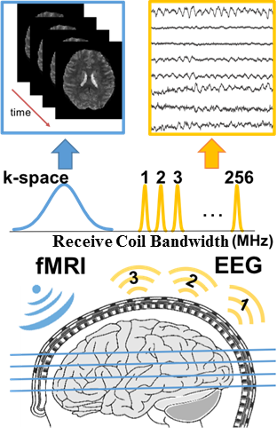
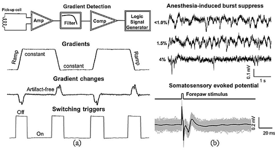
The MR-compatible recording system functions through the use of novel methods that maximally utilize existing MR-hardware and its electromagnetic environment for both synchronization and wireless transmission of non-MR data (Fig. 1). The system is designed to operate in either bipolar or unipolar configuration. The active components combined with minimized cable length and differential signal transmission cancel out electromagnetic interference, while reducing noise and compensating for the signal attenuation. Gradient and RF pulse detection circuit, including on-board pickup coil, amplification, and filtering circuit, reliably detect the ramp periods of the trapezoidal gradients and RF excitation duration to produce logic signals that indicate the electromagnetically silent periods. Utilizing these logical signals stepping stone sampling1 was implemented. Effect of the RF interference and gradient magnetic field changes was minimized by blocking input signals using high fidelity single-pole-double-throw (SPDT) analog switching circuits. High resolution (0.5µV), synchronized digitization circuit (16-bit ADC) provided necessary dynamic range (33mV) while maintaining very low power consumption. An ultra-low power transmitting module wirelessly transmits the digitized EEG signals through FSK demodulation at discrete non-overlapping RF frequencies. Surplus bandwidth of receiver coil, availed by increasing the imaging field of view (FOV) in the readout direction2, is used to accommodate these non-MR signals (Fig.3.b). Upon reception by MR coils, the RF signal is demodulated and separated into EEG signals in one frequency range and MRI data in a different range. The transmitted data is then extracted through standard FSK demodulation algorithm and a locally developed software.Results
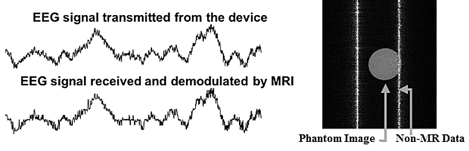
Figure 2(a) shows wireless gradient detection using on-board pickup coil and corresponding switching triggers. A simple illustration of active sensing and subsequent wireless transmission of rat LFP is shown in Figure 2(b). Anesthesia induced burst suppression of spontaneous rat LFP is demonstrated in Figure 2(b)(Top). As concentration isoflurane is increased, resulting in deeper anesthesia, spontaneous LFP progressively transformed towards burst suppression. Fore-paw monophasic current stimulus (1mA) of 2ms duration at 5Hz, generated evoked LFP response in somatosensory cortex is recorded through the device wirelessly (Fig.2.b, bottom). Figure 3(a) displays reliability of extracted EEG signal demodulated from MR raw data compared with original transmitted data. Single channel FSK demodulation of EEG signal within the MR image is demonstrated in Figure 3(b) as non-MR data appears as stripes in besides the original phantom image.Conclusion
The MR-integrated system provides a simple solution for high fidelity neural recording in simultaneous fMRI studies by utilizing MR-surplus hardware. Gradient triggered sampling and analog switching circuit combined with wireless reception of EEG signal by MR coil address technical challenges regarding the signal integrity and electromagnetic artifacts, while reducing the overall complexity by removing the dependence on bulky synchronization and shielding systems. Additionally, further refinement and implementation of the system will be focused on future human applications. Therefore, the success of the current research is expected to open new avenues for widely accessible, integrative neuroimaging tools.Acknowledgements
No acknowledgement found.References
1. K. Anami et al., “Stepping stone sampling for retrieving artifact-free electroencephalogram during functional magnetic resonance imaging,” Neuroimage, vol. 19, no. 2, pp. 281–295, 2003.
2. L. G. Hanson, T. E. Lund, and C. G. Hanson, “Encoding of electrophysiology and other signals in MR images,” J. Magn. Reson. Imaging, vol. 25, no. 5, pp. 1059–1066, 2007.
Figures