0747
Improved Image Quality and Decreased Power Deposition in the Spine at 3T using Extremely High Permittivity Materials1Radiology, Leiden University Medical Center, Leiden, Netherlands, 2Philips Research Hamburg, Germany
Synopsis
High field MR imaging of the spine suffers from a low transmit efficiency. The aim of this study is to improve the transmit profile of the 3T body coil in the spine region by using extremely high permittivity ceramics. This dielectric shimming approach with novel materials offers the opportunity to improve clinical spine image quality on MR systems that are not equipped with multi-transmit hardware. The developed approach is also compared with RF shimming in terms of image quality and power requirements.
Introduction
The spine is a challenging area to image due to its size and elongated shape1. At 3T the signal-to-noise ratio (SNR) and image contrast in the spine area are limited due to a low transmit efficiency. RF shimming is the standard way to try to increase the low intensity regions in the transmit field, but the accompanying costs of the parallel transmit technology mean that many clinical sites are still working with 3T single-channel transmit systems. Therefore, our aim is to explore the feasibility of an alternative to 3T parallel transmit that leads to a similar improvement in transmit field intensity in the spine region, allowing an inexpensive “upgrade” of the scanner hardware. In addition we compare the power efficiency, and hence SAR implications, of the dielectric approach versus RF shimming. In previous studies dielectric pads have shown to increase the transmit efficiency, reduce the peak SAR and decrease the average power in cardiac2, abdominal3,4 and cervical spine5 applications at 3T. In the current work, novel materials6 with permittivity values over three times as high as the permittivities of the dielectric pads are investigated. The design process and safety analysis are performed by simulation and in vivo experiments show the feasibility of this alternative approach.Methods
Dielectric Materials: Rectangular shaped solid blocks made of the piezoelectric material lead zirconate titanate (TRS Technologies, Inc, State College, PA) with dimensions 7x2.5x9 cm3 and a weight of 1.3 kg each were used as dielectric ceramics. The permittivity and the conductivity were measured with a network analyzer (Agilent Technologies, E5061A), $$$\epsilon_r$$$=1075 and $$$\sigma$$$=0.38 S/m . The blocks were positioned in a 7x1 chain with 10 mm air gaps between successive blocks and copper strips connecting all blocks.
EM Simulations: 3D Electromagnetic simulations were performed using the XFdtd EM Simulation Software version 7.4 (REMCOM, State College, PA). An ideal 16-rung body coil driven in quadrature mode was modeled with the male body model "Duke" from the virtual family7 positioned in the center. A 10 mm air gap, realized by a patient mattress in experiments, was inserted between the body and the dielectrics to optimize RF penetration depth. Resulting B1+ and SAR distributions were analyzed.
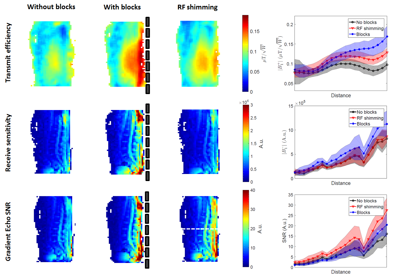
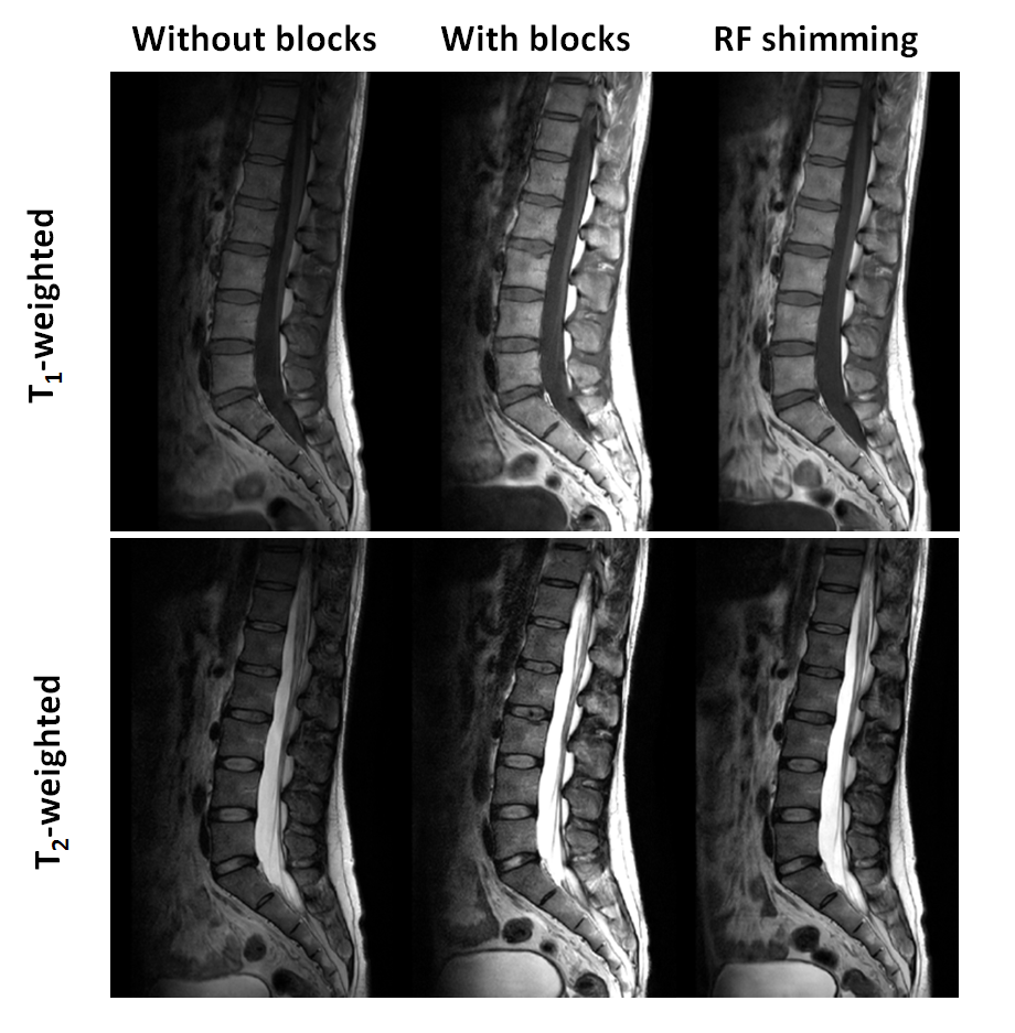
MR Data Acquisition: An Ingenia 3T dual transmit MR system (Philips Healthcare, Best, The Netherlands) was used to acquire in vivo data in ten healthy volunteers. The body coil was used for RF transmission while the signal was received by the table-integrated 12 element posterior coil. A leg cushion was used to bring the back in a flat position, in this way minimizing the distance between the posterior coil and the spine. Quadrature excitation, to mimic a single-channel system, was compared with and without blocks, and also with RF shimming (no blocks). T1- and T2-weighted lumbar spine images were obtained using a standard clinical multislice TSE sequence (voxel size 0.8x1 mm2, slice thickness 4 mm and TE/TR/TSE factor 8ms/641ms/6 and 110ms/4687ms/30, respectively, except for a TR of 705/5155 ms for the T1/T2 weighted RF shimmed scans). B1+ maps were acquired using DREAM8 and SNR and B1- maps were constructed from 1 degree tip angle gradient echo images using the mean of the background noise distribution for normalization.
Results
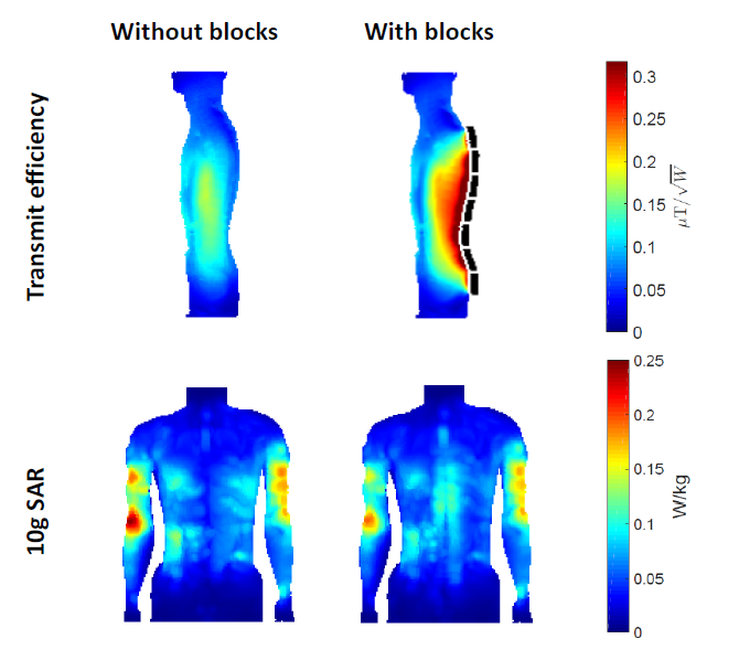
Figure 1 shows the simulated transmit efficiencies and 10g SAR maximum intensity projections without and with the dielectric blocks present. The transmit efficiency in the spine region increases by 75 percent, while the maximum SAR decreases by 20 percent. Experimentally measured and normalized B1+ maps are in agreement with simulation results, and corresponding reconstructed receive sensitivity and SNR maps also show an increase in the spine region (Figure 2). Clinical T1- and T2-weighted images in Figure 3 show a comparable increase in image quality with blocks and RF shimming. However, average input powers and distributions measured for all volunteers (Figure 4) underline an improved power efficiency using the blocks.Discussion and Conclusion
EM simulations and the in vivo data of this study show that the dielectric blocks improve the transmit efficiency in the region of interest. No adverse effects on the receive sensitivity of the array have been found. The locally enhanced receive sensitivity in combination with the focused transmit profile results in higher SNR in the spine region, leading to an increased image quality. In conclusion, the dielectric blocks offer a way to improve image quality in spine imaging if a multi-transmit MR functionality is not available. The dielectric shimming approach also uses less power than conventional RF shimming, indicating an improved power efficiency.Acknowledgements
This project was funded by the European Research Council Advanced Grant 670629 NOMA MRI.References
1. Vertinsky AT, Krasnokutsky MV, Augustin M, et al. Cutting-edge imaging of the spine. Neuroimag Clin N Am 17, 117-136 (2007)
2. Brink WM, Webb AG. High permittivity pads reduce specific absorption rate, improve B1 homogeneity, and increase contrast-to-noise ratio for functional cardiac MRI at 3T. Magn Reson Med. 71, 1632-1640 (2014)
3. Heer de P, et al. Increasing signal homogeneity and image quality in abdominal imaging at 3T with very high permittivity materials. Magn Res Med 68, 1317-1324 (2012)
4. Franklin KM, Dale BM, Merkle EM. Improvement in B1-Inhomogeneity Artifacts in the Abdomen at 3T MR Imaging Using a Radiofrequency Cushion. J Magn Reson Imaging 27, 1443-1447 (2008)
5. Yang QX, Luo W, Rupprecht S, et al. RF Field Enhancement with High Dielectric Constant (HDC) Pads in a Receive Array Coil at 3.0T. J Magn Reson Imaging 38(2), 435-440 (2013)
6. Rupprecht S, et al. Drastic Enhancement and Manipulation of RF Field with Ultra High Dielectric Constant (uHDC) Material at 3T. Proc. Intl. Soc. Mag. Reson. Med. 21 (2013), Cao Z, et al. Dramatic Improvement of Parallel Imaging with High Dielectric Material – Demonstration with Electromagnetic Field Calculations at 123 MHz. Proc. Intl. Soc. Mag. Reson. Med. 21 (2013), Rupprecht S, et al. Signal-to-Noise Ratio Improvement for MR proton spectroscopy at 3T using a ultra High Dielectric Constant (uHDC) Material Sleeve. Proc. Intl. Soc. Mag. Reson. Med. 22 (2014), Sica C, et al. Ultra High Dielectric Constant (uHDC) Head Insert at 3T for Dramatic Reduction of SAR and B1+ inhomogeneity. Proc. Intl. Soc. Mag. Reson. Med. 22 (2014)
7. Christ A, Kainz W, Hahn EG, et al. The virtual family – development of surface-based anatomical models of two adults and two children for dosimetric simulations. Phys. Med. Biol. 55, N23-N38 (2010)
8. Nehrke K, Börnert P. DREAM – a novel approach for robust, ultrafast, multislice B1 mapping. Magn Res Med 68, 1517-1526 (2012)
Figures