0725
Phase II Clinical Hyperpolarized 13C 3D-Dynamic Metabolic Imaging of Prostate Cancer using a B1-insensitive Variable Flip Angle Design1Department of Radiology and Biomedical Imaging, University of California, San Francisco, San Francisco, CA, United States, 2UCSF/UC Berkeley Graduate Program in Bioengineering, University of California, San Francisco, San Francisco, CA, United States, 3Department of Clinical Pharmacy, University of California, San Francisco, San Francisco, CA, United States, 4School of Medicine, University of California, San Francisco, San Francisco, CA, United States
Synopsis
The 3D compressed-sensing echo-planar spectroscopic imaging (CS-EPSI) sequence has enabled 5-dimensional HP-13C imaging of human prostate cancer with full-prostate coverage and high spatial and temporal resolution. A new variable flip angle design substantially desensitizes the sequence to B1 variations for improved quantitative analysis of pyruvate-to-lactate rate constant (kPL) measurements. Pre-clinical animal and clinical patient studies demonstrate that the resulting sequence enabled improved quantitative accuracy while maintaining excellent metabolite SNR.
Purpose
The 3D compressed-sensing echo-planar spectroscopic imaging (CS-EPSI) sequence enables 5-dimensional (3-spatial, 1-temporal, 1 spectral) dynamic HP-13C imaging of human prostate cancer with high spatiotemporal resolution1. A variable flip angle scheme has been applied to improve the metabolite SNR, but the quantitative accuracy of pyruvate-to-lactate conversion can potentially be affected by B1+ inhomogeneity (ΔB1+) with this scheme. Calibration of B1+ is challenging due to the limited natural abundance of 13C in vivo, and can also vary across larger imaging volumes. In this study a new variable flip angle scheme was developed to reduce the sensitivity to ΔB1+ variations across the transmit-coil volume. In vivo HP-13C studies and simulations were conducted to explore the sensitivity of quantitative kPL estimation to ΔB1+, as well as changes in total [1-13C]pyruvate and [1-13C]lactate SNR.Methods
Sequences: The CS-ESPI sequence enables 3D dynamic imaging with high spatiotemporal resolution1. Spectral-spatial excitation pulses for varying flip angles between metabolites were designed using an open-source SS-RF toolbox2. Sequence parameters include TR = 150ms, TE = 4.0ms, spatial resolution: 0.36cm3 (rats), or 0.5cm3 with FOV=9.6x9.6x12.8cm (humans), temporal resolution = 2s, acquisition window = 42s.
MRI experiments: GMP [1-13C]pyruvic acid was polarized in a 5T SpinLab polarizer for 2.5 hours, yielding 35+% polarization of 245mM pyruvate and met all pharmacy specifications prior to injection for the clinical trial study conducted in accordance with the approved IND protocol. A healthy Sprague Dawley rat was used for animal studies.
Simulations: HP-13C pyruvate and lactate signals were generated and fit using a simple and robust two-site exchange model4. B1 inhomogeneity translates into an effective scaling in a given flip angle scheme, denoted by Δθn. Potential variations of ΔB1+= ±20% was assumed for clinical environment.
Results and Discussions
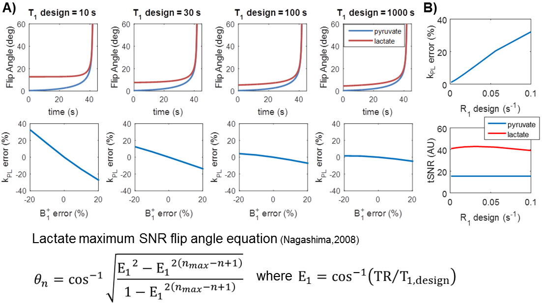
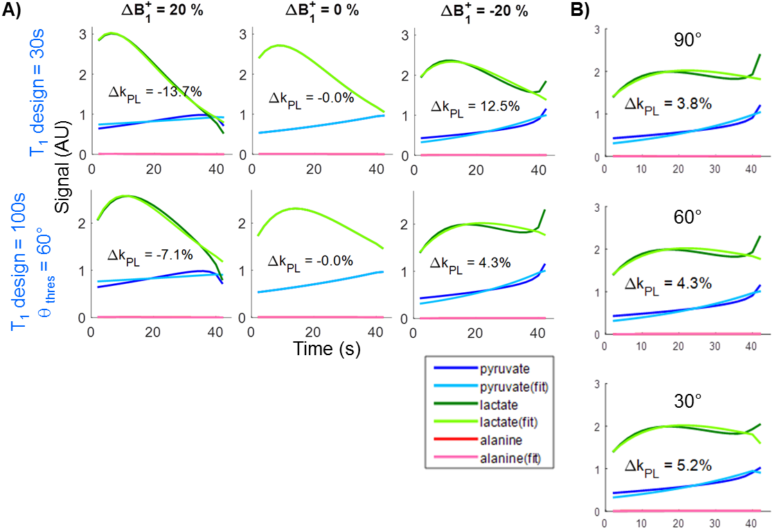
One way of desensitizing the sequence to B1 inhomogeneity is to adjust the effective T1 design factor (T1,design) used to generate the lactate flip angles5. At earlier timepoints, higher amount of magnetization is present, and is thus more susceptible to flip angle variation due to ΔB1+. Increasing T1,design reduces the flip angle for early timepoints (Fig.1A), thereby making the design less sensitive to ΔB1+. Deviating from the “optimal” design parameters comes at the cost of reduced total lactate SNR. Simulations suggest that while the original T1,design =30(s) generates maximum signal, a choice of T1,design =100(s) significantly reduces B1-sensitivitiy (Fig.2A, ΔkPL from 12.5% to 4.3% for ΔB1+= -20%, 13.7% to 7.1% for ΔB1+= 20%) without substantially reducing SNR (Fig.1B, 2.5% lactate tSNR loss).
Additionally, preventing the final flip angle from increasing to 90° helps to mitigate slice profile artifacts6. Such thresholding does not have a significant impact on kPL errors in terms of B1 inhomogeneity, and a 60° threshold was selected based on past simulations6 (Fig.2B). The resulting new spectral-spatial RF pulses using this variable flip angle scheme were 4.0 (ms) long with peak B1 of 0.492(G), 37% shorter and 17% reduced in peak power than our previous clinical design1.
HP-13C imaging of rat kidney measured substantially lower ΔkPL using the new flip angle scheme optimized for B1 insensitivity compared to a previous scheme (T1,design =30(s)) in the presence of ΔB1+ (Fig.3, ΔkPL = 22% vs 63% with ΔB1+= -20%), which agrees with simulation.
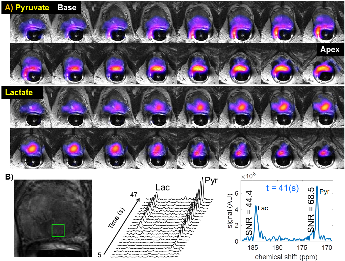
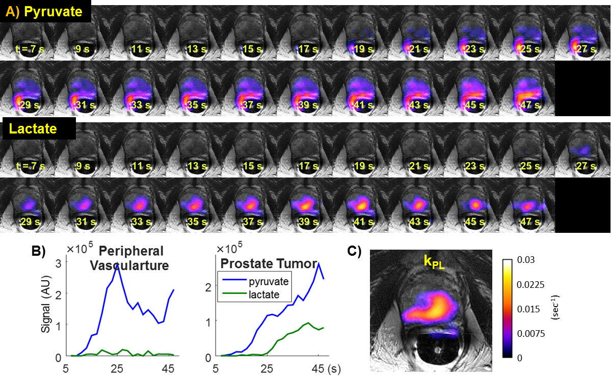
For human studies, the dynamic 3D CS-EPSI acquisition with the improved variable flip angle scheme enabled coverage of the entire prostate gland with good spatiotemporal resolution and high SNR (Fig.4A&5A) with spectroscopic information (Fig.4B). SNR was 68.5 for pyruvate and 44.4 for lactate 41 seconds from injection. The new variable flip angle scheme not only retained excellent pyruvate and lactate SNR, but also preserved the signal timing characteristics compared to our prior studies1. The location of high kPL (Fig.5C) corresponded with a large volume of biopsy-proven Gleason 4+5, 4+4 and 4+3 cancer involving a majority of the left lobe of the prostate and extending into the right peripheral zone.
Conclusions
The 3D CS-EPSI sequence with an improved variable flip angle scheme enabled quantitative analysis of pyruvate-to-lactate conversion with far less sensitivity to B1+ variations. Moreover, the clinical research studies demonstrated the potential to acquire new information in patient exams while preserving the desirable SNR efficiency of the previous design. Improved ΔB1+ insensitivity is beneficial for all HP-13C applications, where calibration of this field is challenging due to the limited natural abundance of 13C in vivo. Furthermore, the proposed variable flip angle schemes will be particularly valuable for human studies over large volumes, e.g. brain and liver, where substantial ΔB1+ will likely be present across the FOV.Acknowledgements
The authors acknowledge funding from R01EB017449, R01EB013427, and P41EB013598.References
[1] Chen H-Y et al., “3D Dynamic Hyperpolarized 13C-Pyruvate MR Metabolic Imaging of Human Prostate Cancer”, ISMRM. 2016.
[2] Larson PE, et al., “Investigation of tumor hyperpolarized [1-13C]-pyruvate dynamics using time-resolved multiband RF excitation echo-planar MRSI,” MRM. 2010; 63(3):582-591.
[3] Nelson SJ et al., “Metabolic imaging of patients with prostate cancer using hyperpolarized [1-13C]pyruvate,” Sci Transl Med. 2013;5:198ra108
[4] Bahrami N et al., “Kinetic and perfusion modeling of hyperpolarized 13C pyruvate and urea in cancer with arbitrary RF flip angles,” Quant Imaging Med Surg. 2014; 4(1): 24-32.
[5] Nagashima K et al., “Optimum pulse flip angles for multi-scan acquisition of hyperpolarized NMR and MRI,” JMR. 2008; 190: 183-188.
[6] Gordon JW et al., “Mis-estimation and bias of hyperpolarized apparent diffusion coefficient measurements due to slice profile effects,” MRM. 2016
Figures