0676
More and faster: multi-timepoint ASL at 150ms time-resolution with whole brain coverage by combining time-encoding, Look-Locker, Multi-Band and flip-angle sweep1C.J. Gorter Center for high field MRI, Department of Radiology, Leiden University Medical Center, Leiden, Netherlands
Synopsis
Multi-timepoint ASL can be acquired with several methods; however, most of these methods have some confounding aspects, such as loss of temporal resolution or limited brain coverage. We combine Hadamard-8 time-encoded-pCASL with Look-Locker readout (4 phases×150ms) for a high temporal resolution; moreover, multiband-factor 4 is used for a time efficient acquisition manner enabling whole brain coverage. This combination of techniques results in 25 PLDs for 16 slices, which provides information about the inflow of labeled blood and tissue perfusion. These images have a temporal resolution of 150ms, whereas inflow of label is measured at 75ms resolution.
Purpose
Multi-timepoint arterial spin labeling (ASL) has been shown to be a powerful tool, especially for circumvention of transit times artefacts as well as to gain detailed insight into the cerebral hemodynamic status1,2,3. A Look-Locker (LL) readout is frequently employed to acquire multi-timepoint data. However, due to the need of dense sampling of the dynamic ASL signal, the coverage of the acquisition is typically limited to 5-7 slices. On the other hand, several separate ASL-scans with different post label delays (PLD) can be acquired. But this leads to a limited temporal resolution, typically a few hundreds milliseconds4,5. The recent introduction of simultaneous multi-slice (SMS, also named multiband (MB)) and time-encoded (te, or Hadamard-encoded) pCASL resulted in new opportunities for high temporal resolution ASL without sacrificing whole brain coverage. The goal of the current study was to achieve the highest temporal resolution multi-PLD pseudo-continuous ASL (pCASL) sequence, while maintaining whole brain coverage. To this end, we will combine SMS, te-pCASL, as well as a LL readout with flip angle sweep within a single sequence, while keeping the temporal resolution at 150ms.Methods
Four healthy volunteers (age 23-58 y.o., 2 female) were scanned with te-pCASL combined with LL acquisition and MB readout using the 32-channel head coil on a 3T scanner (Ingenia, Philips, The Netherlands). All volunteers provided informed consent and the study was approved by the local IRB.
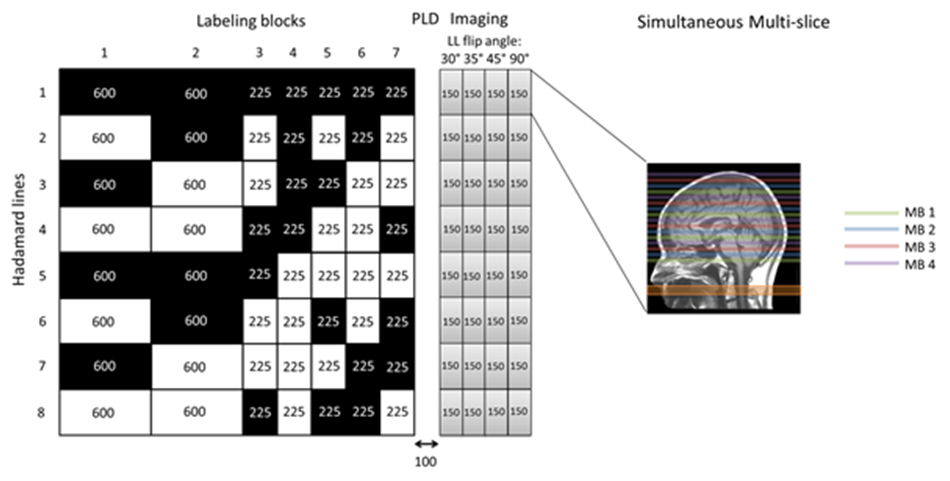
te-pCASL: A Hadamard-8 matrix was used to dynamically encode the pCASL labeling train providing the first approach to obtain dynamic ASL signal (Figure 1). After subtraction, perfusion images with different temporal information are obtained with similar SNR compared to separate pCASL scans2. The total label duration of the te-pCASL labeling was 2325 ms. For background suppression 2 FOCI pulses were applied at 1060 and 2020ms.
Look-Locker: By employing a Look-Locker readout, four multiple images are acquired at an interval of 150ms for each Hadamard line, providing the second source of temporal resolution to the sequence (Figure 1). A flip angle sweep of 30°, 35°, 45° and 90° was applied to maintain a constant signal in the LL readout over the four phases.
Simultaneous multi-slice: To be able to obtain whole brain coverage in the short LL-interval of 150ms, four slices were excited simultaneously with multi-banded radiofrequency (RF) pulses6 in combination with SENSE-factor 1.9 and 0.7 partial Fourier imaging resulting in a total coverage of 16 slices and a voxel-size of 3×3×8mm.
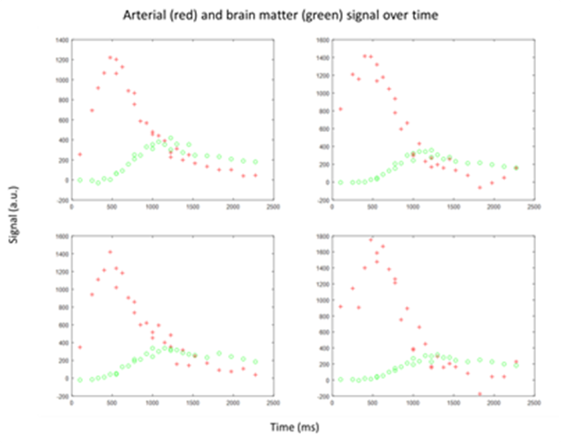
Other MRI parameters: TR/TE=3100/9.52ms and fat suppression=SPIR. In total 192 images (24×8) were acquired in 10:20 min. Post processing: After subtraction according to the appropriate Hadamard scheme, the ASL signal was corrected for the bolus duration and T1 decay; furthermore the ASL maps were ordered chronologically. The combination of the labeling blocks in te-pCASL and the interval of the LL led to interleaved timing of the images for the short PLDs. In addition, the arterial and brain matter signal was studied over time.
Results
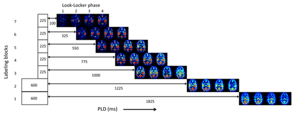
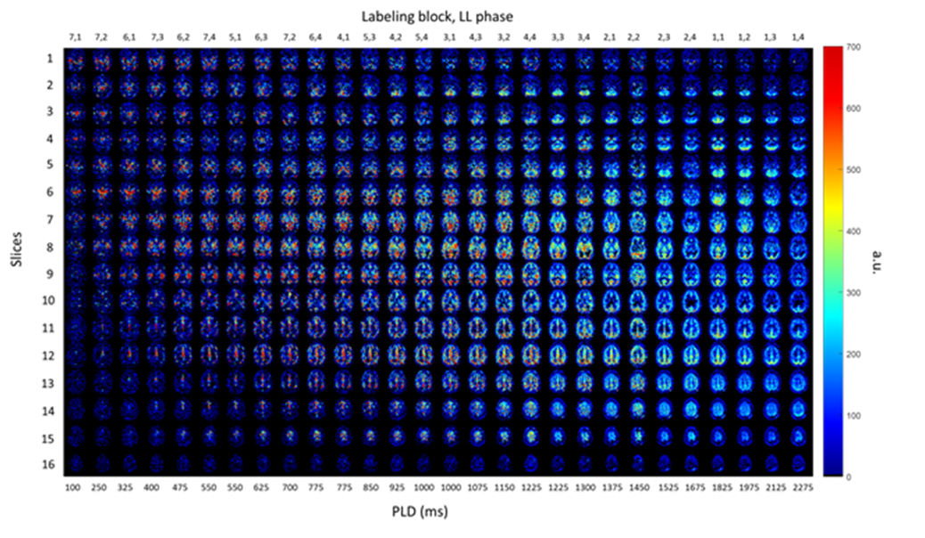
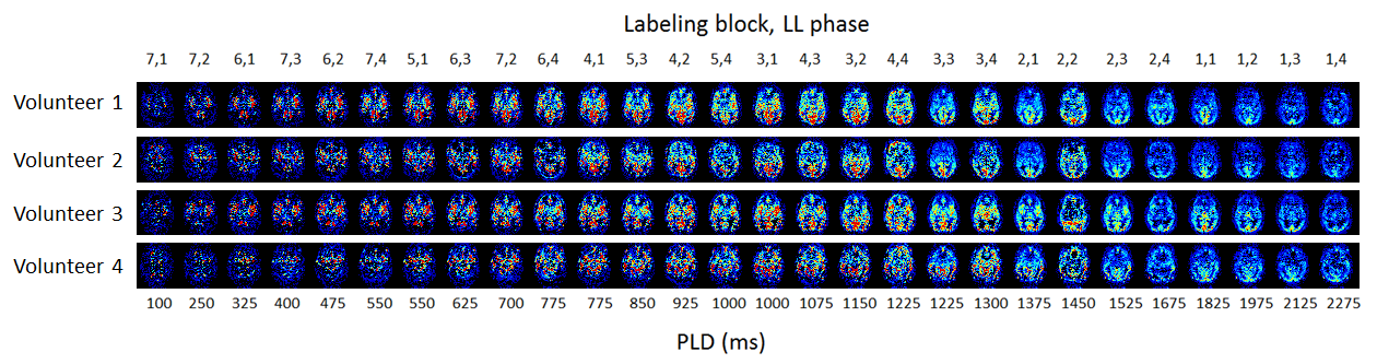
By combining te-pCASL with LL readout, multiple time points are acquired for each Hadamard block (Figure 2), for every slice, 28 images are obtained. The images from different Hadamard blocks with the short PLDs are interleaved, which results in a higher temporal resolution of 75ms to monitor the inflow of labeled spins. Three of these acquisitions have the same bolus duration (225ms) and PLD (550, 775 and 1000ms), but originate from different LL phases. For the longer PLDs a temporal resolution of 150 ms was obtained. Figure 3 shows the complete ASL dataset i.e. 28 chronological time points of 16 slices. Figure 4 shows the 28 chronological time points for a single slice of all the volunteers. The arterial and brain matter signals over time are shown in figure 5.Discussion and conclusion
By combining te-pCASL, SMS readout and LL-acquisition with flip-angle sweep, high temporal resolution, multi-PLD, can be acquired with whole brain coverage in a time efficient manner. The high temporal resolution allows precise fitting of the arrival of the arterial signal and CBF. It was investigated whether a higher temporal resolution could be achieved by exclusion of the fat-suppression pulses. Although, without the SPIR-pulses a 130ms temporal resolution could be achieved, unfolding MB-artefacts became apparent, which were deemed too disruptive. The interleaved acquisitions led to images with the same PLD, but acquired with different LL phase, and some signal differences could be observed. This can most probably be attributed to B1-inhomogeneities and the use of a B1-map should be considered to correct for this.Acknowledgements
No acknowledgement found.References
1. Weiying Dai, Ajit Shankaranarayanan, and David C. Alsop. Volumetric measurement of perfusion and arterial transit delay using hadamard encoded continuous arterial spin labelling. Magn. Reson. Med. 2013;69:1014-1022.
2. Wouter M. Teeuwisse, Sophie Schmid, Eidrees Ghariq, Ilya M. Veer, and Matthias J.P. van Osch. Time-Encoded pseudocontinuous arterial spin labeling: basic properties and timing strategies for human applications. Magn Reson Med, 2014; 72:1712-1722.
3. In vivo hadamard encoded continuous arterial spin labeling (H-CASL). Jack A. Wells, Mark F. Lythgoe, David G. Gadian, Roger J. Ordidge, and David L. Thomas. Magn Reson Med, 2010; 63:1111-1118.
4. Ke Zhang, Seong Dae Yun, and N. Jon Shah. Tripled readout slices in multi time-point pCASL using multiband look-locker EPI. PLoS ONE, 2015; 10(11):e0141108
5. Matthias Günter, Michael Bock, and Lothar R.Schad. Arterial spin labeling in combination with a Look-Locker sampling strategy: inflow turbo-sampling EPI-FAIR (ITS-FAIR). Magn Reson Med. 2001; 46:974-984.
6. Xiufeng Li, Dingxin Wang, Edward J. Auerbach, Steen Moeller, Kamil Ugurbil, Gregory J. Metzger. Theoretical and experimental evaluation of multi-band EPI for high-resolution whole brain pCASL Imaging. NeuroImage. 2015;106:170-181.
Figures