0666
Quantitative texture feature to predict Microscopic portal vein invasion of Hepatocellular carcinoma with contrast-enhanced MR images1Shenzhen Institutes of Advanced Technology, Chinese Academy of Sciences, Shenzhen, People's Republic of China, 2Department of Pathology, Guangdong General Hospital, Guangdong Academy of Medical Sciences, 3Department of Radiology, Guangdong General Hospital, Guangdong Academy of Medical Sciences
Synopsis
Portal vein invasion of Hepatocellular carcinoma (HCC) is a well-known prognostic factor for patients after hepatic resection or liver transplant. Typical risk factors of microscopic venous infiltration (MVI) include large tumor size, multifocality, poor histological grade and non-smooth tumor margins. In this work, we adopt a new techniue for quantitative texture feature Gray-level-run-length nonuniformity (GLRLN) to predict MVI of HCC based on the contrast-enhanced magnetic resonance images (CE-MRI). The present study showed that the proposed texture feature GLRLN in the portal vein phase of CE-MRI yielded best performance as compared with typical markers for the prediction of MVI.
Introduction
Microscopic portal vein invasion (MVI) of Hepatocellular carcinoma (HCC) has been shown to be a crucial histopathologic prognostic factor leading to recurrence after liver transplantation or hepatectomy1,2. In the literature, tumor size, tumor grade, smoothness of tumor margin, capsule, peritumoral enhancement, multifocality, apparent diffusion coefficient (ADC) in diffusion-weighted imaging, tumor markers (alpha-fetoprotein, AFP) were reported to be typical findings for preoperative MVI predictor3,4. Recent advances in quantitative radiomics approaches allow for objective and quantitative characterization that could potentially be used for noninvasive prediction for MVI. To this end, we initially investigate the prediction of MVI of HCCs using a quantitative radiomics approach based on contrast-enhanced MR images and extract a quantitative radiomics feature that can achieve optimal prediction of MVI of HCCs.Methods
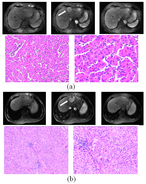
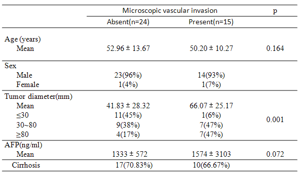
Gd-DTPA CE-MRI data of 39 subjects with resected HCC from July 2012 to October 2015 were consecutively retrieved (Signa Excite HD 3.0T, GE Healthcare, Milwaukee, WI, USA). Demographic and pathological information was summarized in Table 1. Two representative cases of MVI present and absent were shown in Figure 1. Region of interests (ROIs) of HCCs in pre-contrast, arterial and portal vein phases were manually delineated by an experienced radiologist (E.A., 10 years of experience in abdomen radiology) and quantitatively analyzed by the texture feature Gray-level-run-length nonuniformity (GLRLN). The property of GLRLN reflects the degree of nonuniformity of distributed features in four directions (0⁰, 45⁰, 90⁰, 135⁰) and is associated with the texture of vascularity and cellularity. Performance of MVI prediction for the feature was assessed by the alternative free-response receiver operating characteristic (ROC) analysis to predict the presence of MVI and the area under the standard ROC curves (AUC) was also calculated. Optimal feature for MVI prediction was also compared with typical findings illustrated in the literature.Results
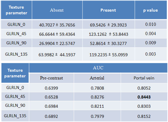
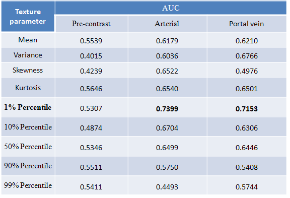
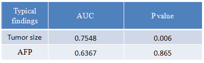
The GLRLN value of HCCs without MVI was significantly smaller than that of the HCCs with MVI, and there were statistical differences of GLRLN in four directions between the absence and presence of MVIs (Table 2). Performance of the texture GLRLN assessed by ROC analysis showed that contrast-enhanced MR images in portal vein phase are better than the arterial phase for prediction of MVI (Table 2). Performance of GLRLN feature was experimentally verified to be better than those based on histogram-based features, tumor size and AFP reported in the literature (Table 3 and Table 4).Discussion
Our finding suggests that the texture GLRLN appears to be an effective feature for prediction of MVI (AUC>0.8) in portal vein phase of contrast-enhanced MR images. As contrast-enhanced MR images embed vascularity and cellularity of HCC, the MVI information can be characterized by image features of GLRLN. Our results also showed that portal vein phase seems to be slightly better than the arterial phase images for prediction of MVI , which was consistent with previous findings with the similar hypothesis that MVI mainly affected portal vein perfusion of tumors in addition to heterogeneous vascularization, cell density, necrosis and fibrosis of a tumor5. Previous researches reported that tumor size, AFP, or histogram–based features were critical factors for prediction of MVI 2,3,5. However, GLRLN feature in the present work is experimentally verified to be better than the performance of histogram-based features, tumor size and AFP.Conclusion
Quantitative texture feature GLRLN was shown to be effective to predict MVI of HCCs based on portal vein phase of the contrast-enhanced MRI. GLRLN yielded better results than the previously reported histogram-based features, tumor size and AFP, which might be helpful for patient management in clinical practice.Acknowledgements
This research is partially supported by grants NSFC U1301258, NSFC 61302171, JCYJ20150630114942291 and No 2011S013.References
1. Gouw, et al. Markers for Microvascular Invasion in Hepatocellular Carcinoma: Where Do We Stand? Liver Translation.2011;10:S72-S80.
2. Unal, et al. Microvascular invasion in hepatocellular carcinoma. Diagn Interv Radiol. 2016;22:125-132.
3. T.M. Pawlik, et al. Tumor size predicts vascular invasion and histologic grade: implications for selection of surgical treatment for hepatocellular carcinoma. Liver Transpl. 2005;11:1086-1092.
4. Renzulli, et al. Can Current Preoperative Imaging Be Used to Detect Microvascular Invasion of Hepatocellular Carcinoma? Radiology. 2016; 279(2):432-442.
5. Huang, et al. Value of MR histogram analyses for prediction of microvascular invasion of hepatocellular carcinoma. Medicine.2016;95: 26.
Figures