0651
Simultaneous multi-slice TSE for clinical MR Imaging of lesions in the knee1Radiology, the Third Hospital of Henbei Medical University, Shijiazhuang, People's Republic of China, 2MR Collaboration NE Asia, Siemens Healthcare, Shang Hai, People's Republic of China, 3Siemens Medical Solutions USA, Inc., Minneapolis, MN, United States
Synopsis
To introduce simultaneous multiple slices (SMS) turbo spin echo (TSE) and to evaluate its image quality and diagnostic accuracy for lesions in the knee. Participants were examined by SMS and routine TSE sequences. Both sequences were evaluated by three radiologists with subjective and objective scores in T1- and PD-weighted images. The diagnostic value in lesions was evaluated. SMS requires less scan time and offers similar imaging quality and diagnostic rate compared to routine TSE sequence. SMS is a valuable technique for MR examination of the knee.
Purpose
To introduce simultaneous multiple slices (SMS) 1-4 turbo spin echo (TSE) with gradient-based “controlled aliasing in parallel imaging result in higher acceleration” (CAIPIRINHA) 5, 6 as a fast sequence for musculoskeletal imaging, and to evaluate the image quality and diagnostic accuracy of lesions in the knee.Methods
Ten healthy volunteers and 20 patients, six with osteoarthritis in the knee joint, two with meniscal damage, seven with bone disease around the knee (one osteochondroma of the distal femur; two osteosarcoma; one fibrous histiocytoma; one fibrous dysplasia of bone of the proximal tibia; one bone infarction of femur and tibia; one pigmented villonodular synovitis), three with bone fracture, and two with soft-tissue hemangiomas around the knee joint were included in this study. All subjects were examined using 3T MRI (MAGNETOM Verio, Siemens, Erlangen, Germany) with an 8-channel knee coil or a four-channel flex coil, using a prototype SMS TSE and routine TSE sequences. The basic acquisition parameters for TSE were as follows: FOV = 160×160 mm2, matrix = 320×256, slice thickness = 3 mm, slices number= 36, gap = 10%, flip angle = 150 degree. T1-weighted images were acquired with TE/TR = 13/499 ms, turbo factor = 3; proton-density (PD) images were obtained with TE/TR = 40/3200 ms, turbo factor = 2; For the SMS sequence, slice acceleration factor and FOV shift factor were both two. Sagittal T1-weighted and PD-weighted TSE with fat suppression were used to compare the image quality and reliability. Three radiologists, each with more than 10 years’ experience in musculoskeletal imaging and blinded to the type of sequences, scored images of 10 volunteers based on artifacts, visualization of bone, cartilage, ligament and meniscus using a 5-grade score. The contrast-to-noise ratios (CNRs) of ligament/bone, meniscus/bone and cartilage/bone in the sagittal T1-weighted images were compared between the SMS and routine TSE. The ligament/effusion, meniscus/effusion, cartilage/effusion and bone/effusion in PD-weighted images were compared between the SMS and routine TSE images. The diagnostic value of SMS TSE in knee lesions was evaluated by the same three radiologists based on the size and edge of the lesions, the relationship with the surrounding structures, and the degrees of the injury in all 20 patients.Results and Discussion
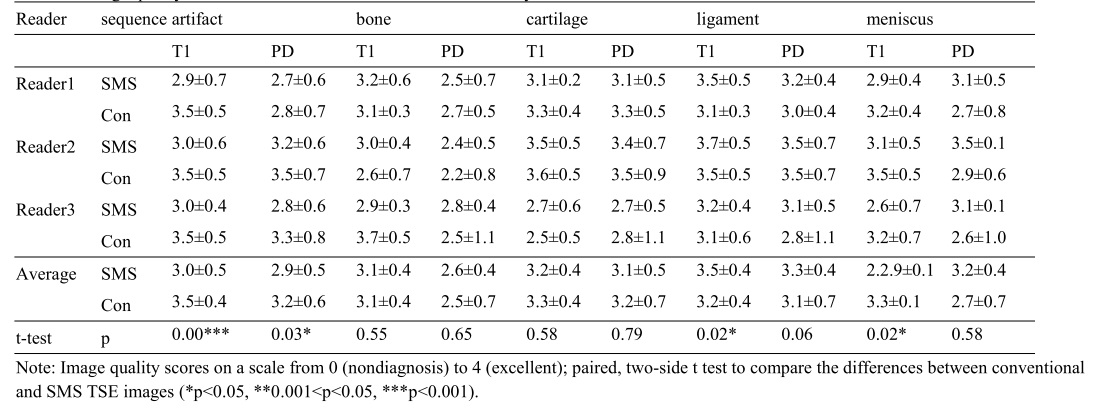
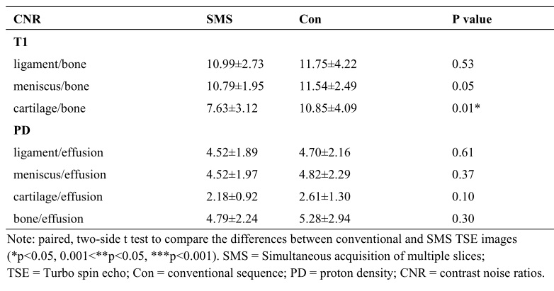
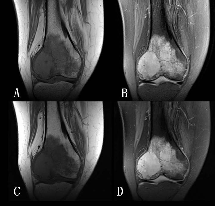
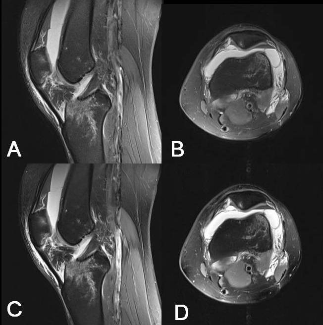
Simultaneous multi-slice techniques significantly decreased the overall scan time. The scan time for SMS TSE was 2 minutes 32 seconds, compared to 5 minutes 4 seconds for the T1-weighted TSE sequence. Similarly, for PD-weighted fat-saturation imaging, SMS TSE scan time was 2 minutes 40 seconds, whereas TSE scan time was 5 minutes 20 seconds. The three readers showed fair (0.38) to good (0.63) (mean ICC: 0.47 ± 0.08) agreement on the visual evaluation results. The summary of evaluation results is shown in Table 1. For the artifacts, routine TSE had significantly higher scores than SMS TSE in both T1- and PD-weighted images. In terms of visualizing structures, SMS and conventional TSE PD-weighted images showed no difference in bone, cartilage, ligaments and meniscus. SMS-TSE T1-weighted images showed higher scores for ligament, no differences for bone and cartilage, and lower scores for meniscus compared to conventional TSE. In terms of CNR in T1-weighted images, SMS and routine TSE of ligament/bone and meniscus/bone showed no difference. SMS TSE had lower CNR than routine TSE images for cartilage/bone. For PD-weighted images, there was no difference in the CNRs between SMS and routine TSE (Table 2). Both sequences showed clear borders in the cases of benign tumors and tumor-like lesions of bone, allowing for the precise visualization of a tumor and its relation to surrounding structures. Figure 1 shows the case of a patient with right-knee pain for 3 months. Atypical fibrous histocytoma was suspected based on MRI, and was confirmed with percutaneous fine needle aspiration biopsy. For musculorskeletal disorders in the knee joint, these two sequences showed the same diagnostic rates. Figure 2 shows a case of left-knee pain 9 hours after injury. There was a tibial plateau fracture with slight increases of peripheral effusion and soft-tissue swelling. The medial retinaculum of the left patella was also injured. Again, the two sequences showed the same diagnostic rates.Conclusion
SMS TSE requires less scan time and offers similar image quality and diagnostic rates compared to routine TSE sequence. This novel sequence is valuable for the examination of the knee, especially for large-coverage imaging with high spatial resolution.Acknowledgements
No acknowledgement found.References
[1]. Gaudiano C, Clementi V, Busato F, et al. Diffusion tensor imaging and tractography of the kidneys: assessment of chronic parenchymal diseases. Eur Radiol. 23(6):1678-85 2013.
[2]. Lu L, Sedor JR, Gulani V, et al. Use of diffusion tensor MRI to identify early changes in diabetic nephropathy. Am J Nephrol. 34(5):476-82 2011.
[3]. L. Chen, A. Vu, J.Xu, etal. Evaluation of Highly Accelerated Simultaneous Multi-Slice EPI for FMRI. Neuroimage. 104:452-459 2015.
[4]. B Orjan A.Gagoski, Berkin Bilgic, Cornelius Eichner, et al. RARE/Turbo Spin Echo Imaging with Simultaneous MultiSlice Wave-CAIPI Mag Reson Med.73(3): 929-938 2015.
[5]. Breuer FA, Blaimer M, Mueller MF, et al. Controlled aliasing in volumetric parallel imaging (2D CAIPIRINHA). Magn Reson Med. 2006;55(3):549-56.
[6]. Breuer FA, Blaimer M, Heidemann RM, Mueller MF, Griswold MA, Jakob PM. Controlled aliasing in parallel imaging results in higher acceleration (CAIPIRINHA) for multi-slice imaging. Magn Reson Med. 2005;53(3):684-91.
Figures