0648
Age Related Differences in Shear Strain in Medial Gastrocnemius: Implications for Lateral Transmission of Force1Physics, UC San Diego, San Diego, CA, United States, 2Physics, San Diego State University, San Diego, CA, United States, 3Radiology, UC San Diego, San Diego, CA, United States
Synopsis
The disproportionate loss of muscle force in comparison to loss of muscle mass with age remains unexplained. Recent studies indicate that the remodeling of the extracellular matrix (ECM) may disrupt lateral force transmission pathways mediated by the ECM. Shear strain is the mechanism that supports lateral transmission of force. We quantified shear strain in muscle from the strain rate tensor derived from velocity encoded phase contrast dynamic images of the in-vivo human calf muscle under isometric contractions. The maximal shear strain was significantly lower in the older cohort compared to the younger cohort which potentially identifies that lateral force transmission decreases with age.
Purpose
Aging results in muscle force loss disproportionate to the loss of muscle mass1. Recent studies indicate that compromised lateral transmission of force pathways could potentially contribute to the force loss with age2. Shear strain in the endomysium has been postulated as the most likely mechanism of lateral force transmission3 and can be derived from strain rate tensors computed from velocity encoded phase contrast images. This study explores the age related shear strain in the medial gastrocnemius (MG) from a series of velocity encoded phase contrast (VE-PC) images acquired under isometric contraction.Methods
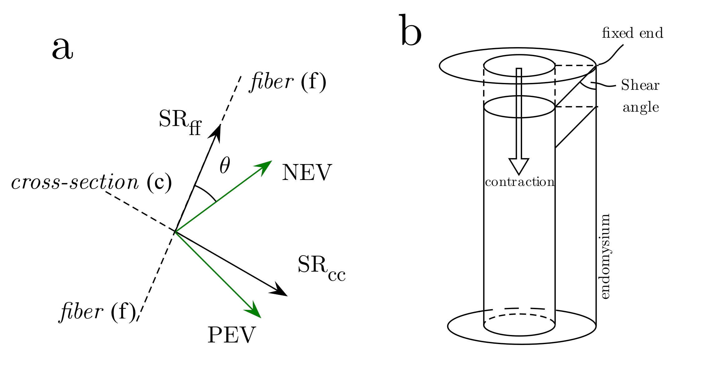
Force measurements and Magnetic Resonance Imaging studies were performed on nine young (27.5 ± 4.8 years, height: 159.6 ± 6.5 cm, mass: 54.6±7.3 kg) and eight senior (77.6 ± 7.3 years, height: 154.3 ± 2.9 cm, mass: 57.9 ± 3.7 kg) female subjects recruited after IRB approval. Dynamic images of the lower leg were acquired during isometric contraction using a gated VE-PC sequence on a 1.5-T GE scanner with a specially designed 8-Ch phased array coil, and the leg in a plaster cast4. Images were velocity encoded in all three directions, and collected in 22 phases, 7 oblique-sagittal contiguous slices, 5mm thick and 1.7x1.7 mm in-plane resolution. Subjects were provided visual feedback within the scanner to maintain consistent contractions at ~40% MVC. Muscle fascicles were manually delineated on water-suppressed images to obtain fiber orientation during the dynamic cycle and tracked using VE-PC. The symmetric part, of the strain rate tensor SR tensor was calculated from the spatial gradient tensor, and diagonalized; eigenvectors corresponding to the positive (PEV) and negative (NEV) eigenvalues were analyzed separately. The SR tensor in the principal axis basis (no off-diagonal terms) was rotated to that of the muscle fiber (fascicle direction) basis (Fig. 1 shows the relationship between the two basis as well as the source of shear strain). Maximum values of shear strain were then obtained by rotating the tensor from the muscle fiber basis by 45o.Results
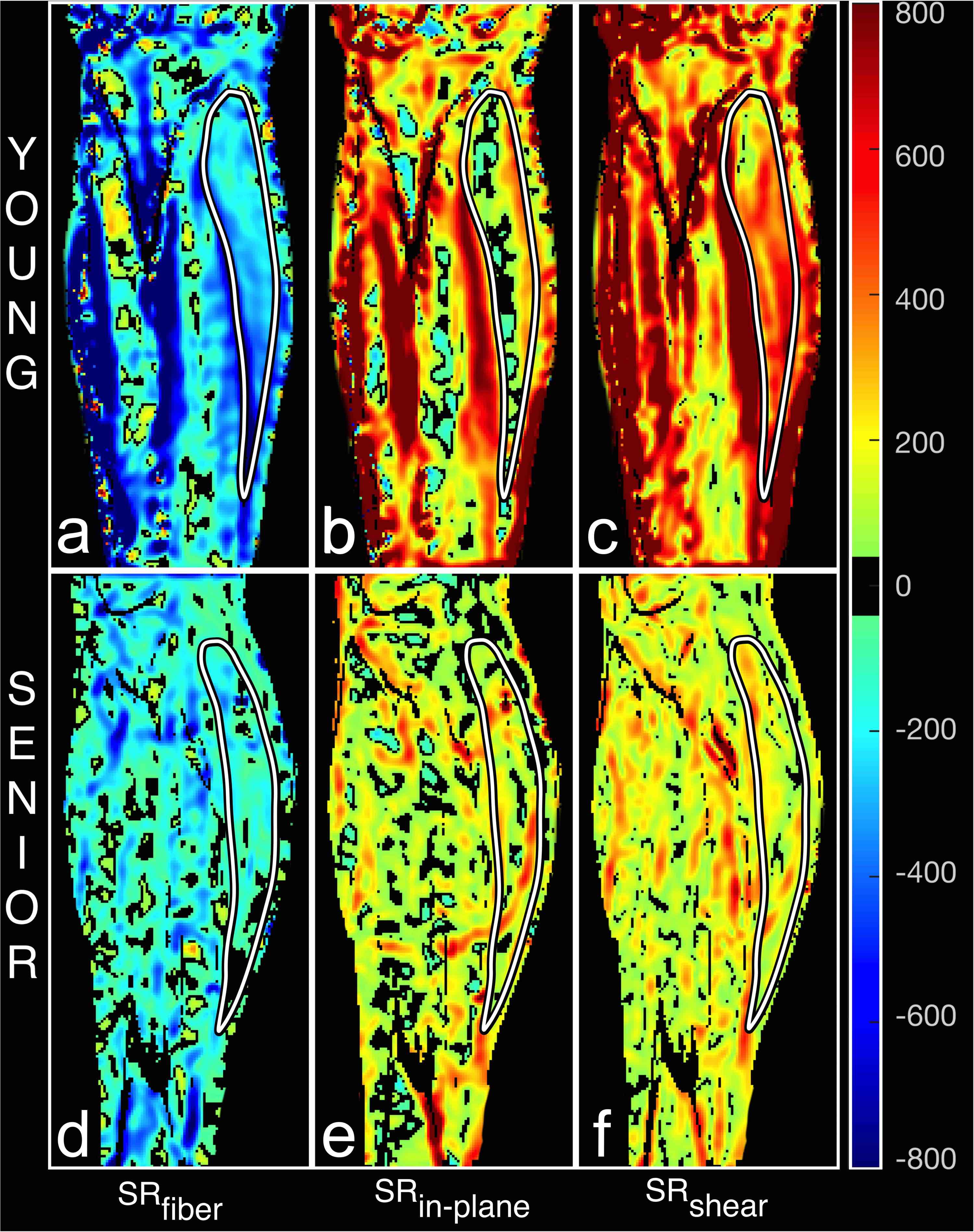
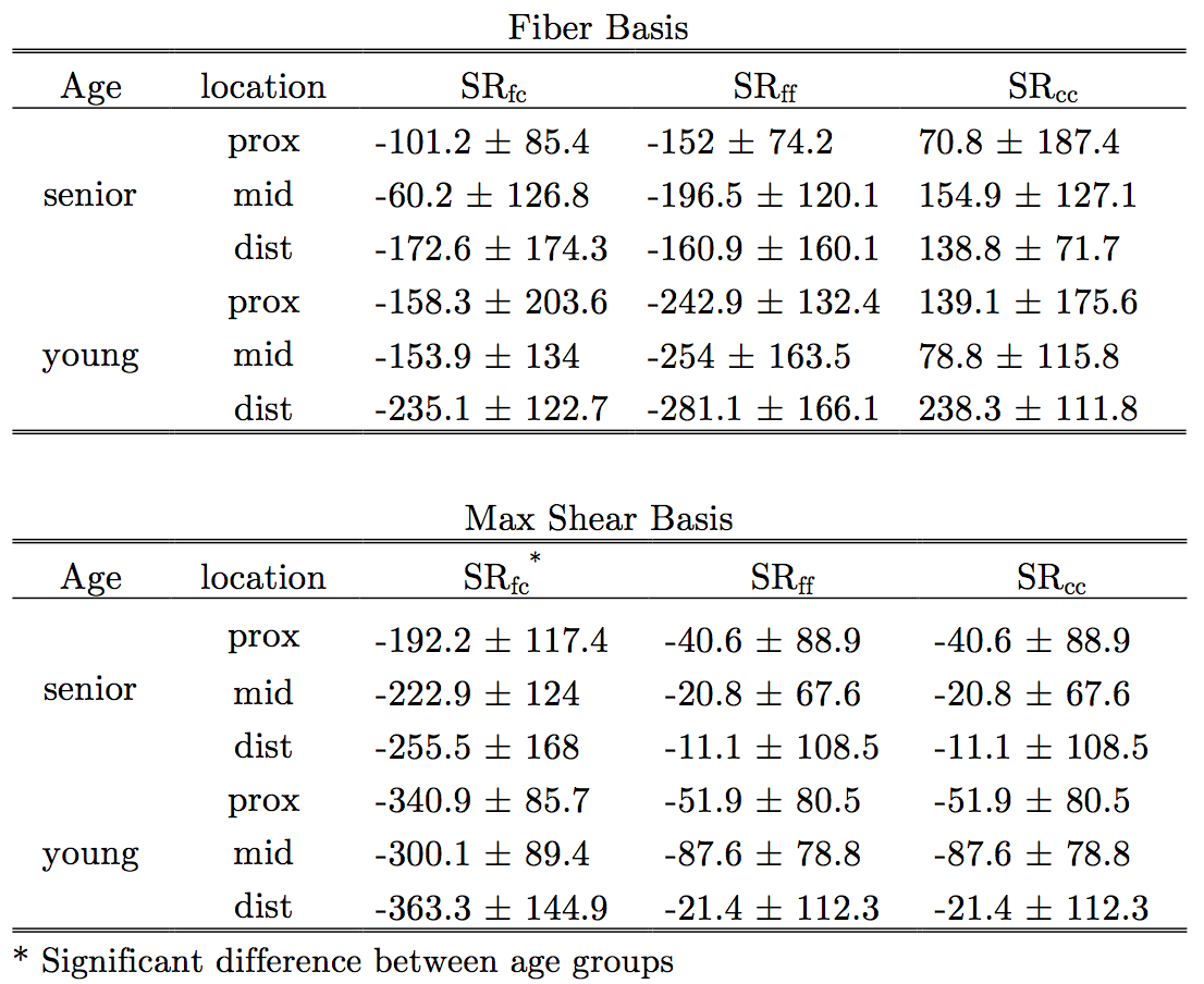
Fig. 2 shows the maps of the strain along the muscle fiber (SRfiber or SRff), in the fiber cross-section (SRin-plane or SRcc) and shear strain in the fiber basis (SRfc) from one young and one old subject at the peak of the contraction cycle. The maps of the shear strain clearly show decreased values in the older subject (Fig. 2). Table 1 lists the shear strain in the muscle fiber basis as well as the maximum shear basis for the ROI placed at proximal, middle, and distal regions. While a decrease in shear strain was seen in the muscle fiber basis, significant differences [F(1,34) = 10.302, P=0.003] were only seen in the maximum shear strain basis.Discussion
Force generated in the muscle fibers is transmitted longitudinally through the myotendinous junction and laterally through myofascial pathways. While the contractile and neural determinants of force loss with age have been studied extensively, very little is known about lateral transmission of force. The lateral transmission of force was shown to decrease in a rodent model of aging and accounts for as much as 50% of the force loss with aging. However, the challenge is that there are no non-invasive approaches to determine lateral transmission of force. This report on shear strain is the first non-invasive marker of lateral force transmission and is a reflection of extracellular matrix (ECM) remodeling that occurs with age. ECM remodeling includes increase in the width of the endomysium and perimysium as well as in the stiffness of the ECM (potentially from abnormal deposition of collagen). The decrease of shear strain with age indicates that some of the force loss in the aging cohort could be attributed to a reduction in lateral force transmission.Conclusions
The potential to determine lateral transmission of force non-invasively offers a powerful tool to monitor the functional consequences of structural remodeling of the ECM and is one that will enable the design of optimum rehabilitative strategies.Acknowledgements
This work was supported by National Institute of Arthritis and Musculo- skeletal and Skin Diseases Grant 5RO1-AR-053343-08.References
[1] Goodpaster BH, Park SW, Harris TB, Kritchevsky SB, Nevitt M, Schwartz AV, et al. J Gerontol A Biol Sci Med Sci. 2006;61(10):1059-64.
[2] Ramaswamy KS, Palmer ML, van der Meulen JH, Renoux A, Kostrominova TY, Michele DE, et al. J Physiol-London. 2011;589(5):1195-208.
[3] Zhong X, Epstein FH, Spottiswoode BS, Helm PA, Blemker SS. J Biomech. 2008;41(3):532-40.
[4] Sinha U, Malis V, Csapo R, Moghadasi A, Kinugasa R, Sinha S. Magn Reson Med. 2015;73(5):1852-1863.
Figures