0638
3D True Polarity Recovery with Independent Phase Estimation Using Multi-layer Stacks Based Region-Growing (3D-TRIPS)1Department of Bioengineering, University of Washington, Seattle, WA, United States, 2Department of Radiology, University of Washington, Seattle, WA, United States
Synopsis
3D whole heart phase sensitive late gadolinium enhanced (LGE) imaging can improve myocardial scar detection. Phase sensitive inversion recovery (PSIR) LGE is sub-optimal for clinical 3D application as it doubles the scan time with a fully sampled reference image. To address this, we develop 3D True Polarity Recovery with Independent Phase Estimation Using Multi-layer Stacks Based Region-Growing (3D-TRIPS) for direct reconstruction of 3D phase sensitive images without need for a separate reference scan. We demonstrate that 3D-TRIPS images shows good agreement and less artifacts compared with PSIR images. 3D-TRIPS will allow 3D-LGE imaging with half the scan time of PSIR.
Purpose
Phase sensitive inversion recovery (PSIR)1 is a commonly used clinical tool for late gadolinium enhancement (LGE) myocardial infarction detection due in large part to its robustness to poorly chosen inversion times. In order to restore the true polarity of inversion recovery images, however, PSIR requires a fully sampled reference image to remove the background phase of the inversion recovery image. This effectively doubles the scan time and decreases its clinical applicability for 3D whole heart imaging. In this study, a method termed 3D True Polarity Recovery with Independent Phase Estimation Using Multi-layer Stacks based Region-growing (3D-TRIPS) was developed to directly reconstruct 3D phase sensitive images without the phase reference image.Method
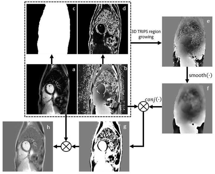
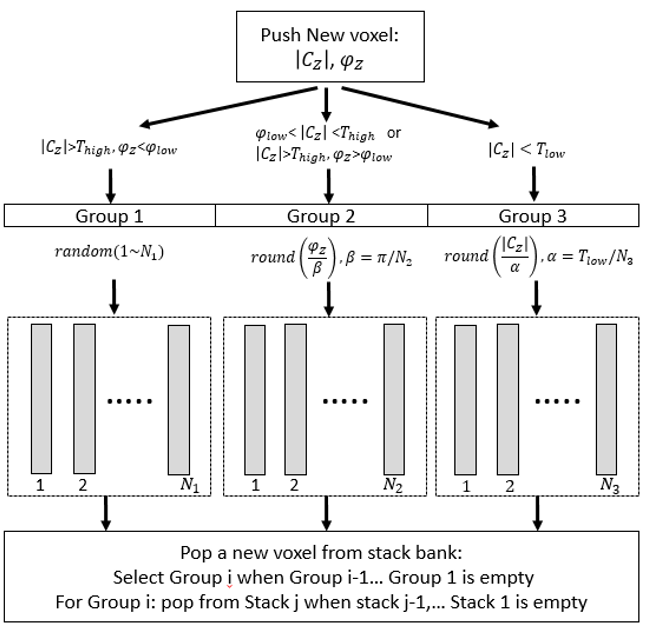
3D-TRIPS algorithm: A flow chart of 3D-TRIPS is shown in Figure 1. The basis for 3D-TRIPS is that the background phase map is specially smooth2 . Therefore, if the difference between the phase of a voxel(φz)and the average of its neighboring voxels’ background phase is smaller than π/2, is regarded as the background phase (φbackground), otherwise φbackgroundis set to π-φz. The phase sensitive image is reconstructed after removing from . Region-growing method is used with 3D-TRIPS to traverse the voxels of interest2. However, an inappropriate region-growing track can be sensitive to large regions of error caused by the error of a few voxels. To quickly and robustly extract a 3D background phase map, a structure with three layers of ordered stacks was developed to direct the region-growing track (Figure 2). Supported by Monte-Carlo simulation, this structure divides the voxels into three groups based on their reliability for background phase extraction: Group 1 includes voxels with high SNR and small neighboring phase difference; Group 2 includes voxels with moderate SNR or high SNR with large neighboring phase difference; Group 3 includes low SNR voxels. Each group is stored in a layer of stacks and within each layer, the stacks are sorted based on the range of SNR or neighboring phase difference they are assigned to store. During region-growing, the voxels from the non-empty stacks with the highest priority are processed first, guaranteeing that each voxel relies on the neighboring voxels with similar or better quality to estimate the background phase. A large number of stacks within each layer promotes the even distribution of growing tracks.
MRI scan and evaluation: To validate 3D-TRIPS, 15 patients undergoing cardiac MR were recruited and scanned on a 3T scanner (Philips Ingenia, the Netherlands) using dStream anterior and posterior coils. The scan parameters for 3D PSIR: 2RR interval [with reference phase image acquired in second RR], TR/TE 5.2/2.5ms, flip angle 25o, FOV 350×350×125mm3, resolution 1.1×1.1mm², slice thickness 5mm, scan plane cardiac short axis, respiratory navigator gating. Real and imaginary images were saved prior to phase correction. PSIR images were acquired 5-10 minutes after the injection of contrast. All study procedures were approved by the local institutional review board. The images were evaluated by the metric Sindex 2 and reviewed by two blinded radiologists. Sindex is defined as Ncorrect/Ntotal , where Ncorrect represents the number of voxels with same polarity between 3D-TRIPS images and PSIR images, while Ntotal represents the total number of voxels excluding the background and regions of low MRI signal area such as the lung.
Results and discussion
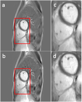
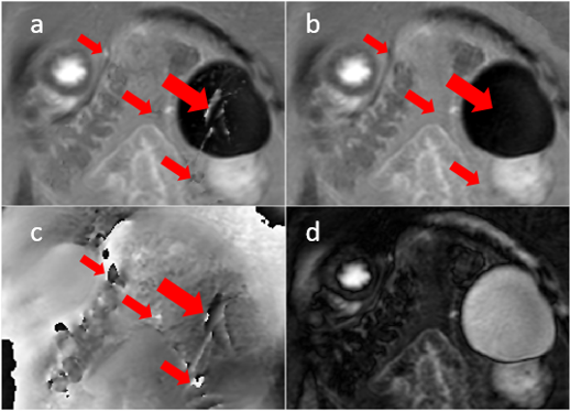
The proposed algorithm was successfully applied to the 3D datasets for all 15 patients, with processing time 4 – 6 minutes. Images reconstructed with 3D-TRIPS showed good overall agreement with PSIR images (Figure 3). The mean Sindex of the 15 patients was0.950± 0.022, with a range of [0.900, 0.980]. Compared with PSIR images, 3D-TRIPS images are free from artifacts often introduced by the reference images. These artifacts can be caused by the motion, flow and eddy currents in the reference image, as well as misregistration between the inversion recovery and reference images (Figure. 4). Subjective review by the two blinded readers showed that 3D TRIPS was often favored over PSIR based on reduced myocardial swaps (53% vs. 3%, p=0.001), swaps outside the heart (70% vs. 17%, p=0.010), and blurring (57% vs. 7%, p=0.007). The details and analysis of the blinded review is summarized in another abstract submitted to this meeting.Conclusion
By application of the three-layer stacks structure, 3D-TRIPS can directly reconstruct 3D phase sensitive LGE images of the whole heart with better image quality. Use of 3D-TRIPS will enable 3D LGE with only half the acquisition time as compared with conventional PSIR. This method has potential to make 3D myocardial phase sensitive imaging more feasible for clinical application.Acknowledgements
NIH 1R21EB017514-01A1 and Philips HealthcareReferences
[1] Kellman, Peter, et al. "Phase-sensitive inversion recovery for detecting myocardial infarction using gadolinium-delayed hyperenhancement." Magnetic resonance in medicine 47.2 (2002): 372-383. [2] Wang, Jinnan, et al. "Referenceless Acquisition of Phase-sensitive Inversion-recovery with Decisive reconstruction (RAPID) imaging." Magnetic resonance in medicine 72.3 (2014): 806-815.Figures