0619
Glial activation measured by [11C]-PBR28 PET correlates with 1H-MRS brain metabolites in amyotrophic lateral sclerosis1Radiology, A. A. Martinos Center for Biomedical Imaging, Massachusetts General Hospital, Charlestown, MA, United States, 2Harvard Medical School, 3Neurology, Neurological Clinical Research Institute (NCRI), A. A. Martinos Center for Biomedical Imaging, Massachusetts General Hospital, Boston, MA, United States
Synopsis
The purpose of our study was to evaluate the relationship between glial activation assessed by [11C]-PBR28 positron emission tomography, and neuronal integrity and gliosis/neuroinflammation measured by magnetic resonance spectroscopy in people with amyotrophic lateral sclerosis (ALS). Glial activation measured by increased [11C]-PBR28 uptake correlated with increased levels of myo-Inositol/Creatine, a spectroscopic marker of gliosis/neuroinflammation in the brain stem and motor cortices. Furthermore, increased [11C]-PBR28 uptake correlated with neuronal damage measured by decreased N-acetylaspartate/Creatine levels. To our knowledge, this is the first study to evaluate the relationship between glial activation, measured by [11C]-PBR28 PET, and brain metabolites assessed by MRS.
Purpose
The purpose of our study was to evaluate the relationship between glial activation assessed by [11C]-PBR28 positron emission tomography (PET), and neuronal integrity and gliosis/neuroinflammation measured by proton magnetic resonance spectroscopy (1H-MRS) in people with amyotrophic lateral sclerosis (ALS). There is a vast body of literature using advanced neuroimaging techniques such as 1H-MRS to study in vivo molecular changes in the brain of patients with ALS [1-5]. The majority of these studies have described a reduction in N-acetyl aspartate (NAA) concentrations [6-8] or NAA ratios (e.g. NAA/Creatine or Choline) [9-16] suggesting either neuronal loss or injury in the motor cortex of ALS patients compared to heathy controls. Elevated myo-Inositol or mI/Cr ratios have been detected in the motor cortex in people with ALS [10, 17-19] indicating glial activation / neuroinflammation in the brains of these patients. These studies showed predominantly metabolic alterations in the motor cortices and brain stem [7, 20]. Utilizing PET radiotracers of neuro-inflammation several groups, including ours [21] have provided evidence of increased glial activation in ALS patients, specifically in the motor cortex and brain stem [22-24].Methods
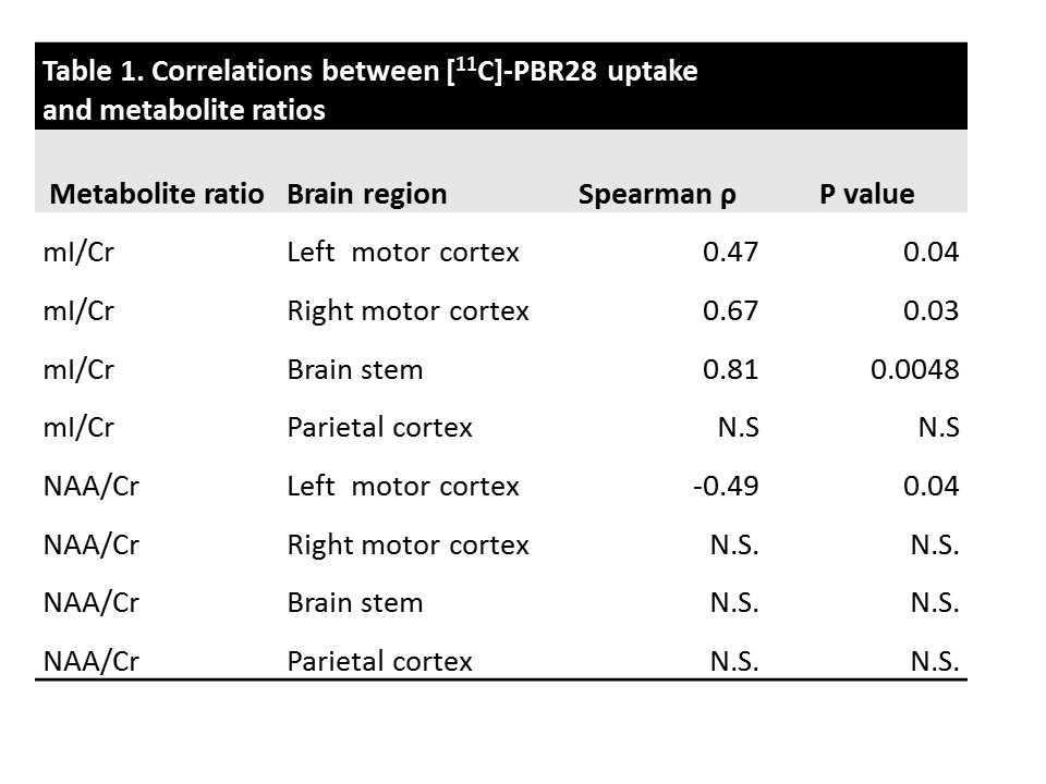
A total of 24 participants with either limb onset ALS (N=18) or bulbar onset ALS N= 6) were included in this study. The participants underwent brain imaging using 3T MR/PET (Siemens Erlangen) imaging using an 8 channel head coil. PET data were collected for 90 minutes post i.v. administration of up to 15 mCi of [11C]-PBR28. PET images were reconstructed using the Ordinary Poisson Ordered Subset Expectation Maximization 3D algorithm. Attenuation map were obtained from the MPRAGE data using an in-house developed method [25]. Standardized Uptake Values (SUV) were obtained voxelwise by normalizing mean tissue radioactivity by injected dose by weight. SUV ratios (SUVR) were then obtained by dividing each voxel SUV by the average SUV from the whole brain [21, 26]. All subjects were genotyped for the Ala147Thr TSPO polymorphism, which explains difference in radioligand binding affinity [27]. Low affinity binders were excluded, and high or mixed affinity binding status was modeled as a covariate in our analyses. Single voxel (2 x 2 x 2 cm3) MRS data were acquired in four regions of the brain using a PRESS sequence with TE/TR 30/1700 ms, and 128 averages: left motor cortex (22 subjects), right motor cortex (17 subjects), brain stem (10 subjects), and parietal cortex as control region (14 subjects). Of note, brain stem and parietal cortex voxels were added later to the study design which explains the lower number of MRS scans in these regions. Furthermore, 3 brain stem voxels were excluded due to suboptimal shimming. LCmodel was used to analyze MRS data and to measure brain metabolite ratios pertaining to neuronal viability (NAA/Cr) and glial activation/inflammation (mI/Cr). Freesurfer’s tools were employed to create 3D volumes with the same spatial extent and location as the MRS Voxels. Pearson correlations were conducted to study the relationship between [11C]-PBR28 SUVR and brain metabolite ratios within each voxel, correcting for TSPO polymorphism.Results
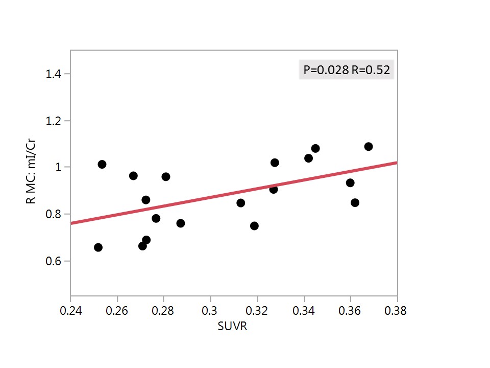
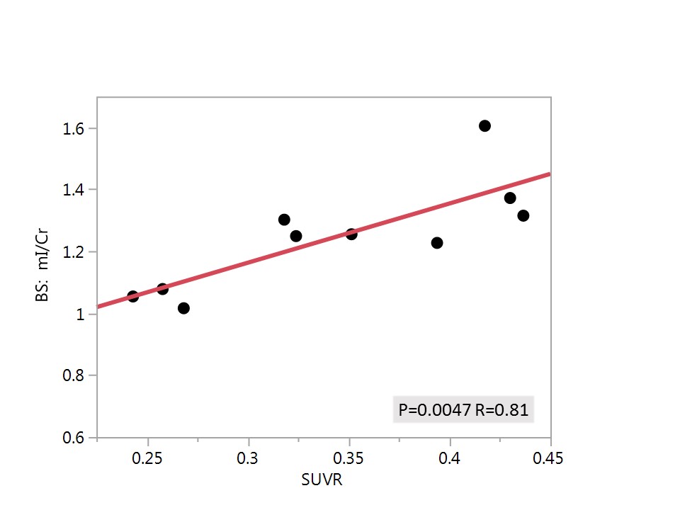
Pearson correlation coefficient revealed an inverse relationship between glial activation, represented by [11C]-PBR28 uptake and NAA/Cr in the left motor cortex (P=0.04 R=-0.49) but not in other brain regions (Figure 1 and Table 1). Positive relationships were found between [11C]-PBR28 uptake and mI/Cr in the left motor cortex (P=0.04 R=0.47), right motor cortex (P=0.03 R=0.67) and the brain stem (P=0.0048 R=0.81) (Figures 2 and 3). No correlations between NAA/Cr or mI/Cr and [11C]- PBR28 uptake were detected in the parietal cortex, which served as control region.Discussion
Our study showed that glial activation represented by increased [11C]-PBR28 uptake correlated with increased mI/Cr measured by MRS. These correlations are detected specifically in the brain stem and left and right motor cortices, both brain regions known to be affected in patients with ALS. To our knowledge, this is the first study to evaluate the relationship between glial activation, measured by [11C]-PBR28 PET, and the proposed spectroscopic marker of inflammation, mI/Cr. Furthermore, our data revealed that glial activation in the left motor cortex measured by PET correlated with neuronal injury or loss, detected by decreased NAA/Cr. Several studies have shown that this region is severely affected in ALS patients. No correlations were found between [11C]- PBR28 uptake and any of the metabolites in the control region (parietal cortex) Clinically there are no signs or symptoms to suggest that the parietal lobes are involved in ALS and prior imaging studies did not show any changes).Conclusion
Combining multiple neuroimaging modalities is of importance to unraveling ALS disease mechanisms.Acknowledgements
The study was conducted at the A. A. Martinos Center for Biomedical Imaging and funded by a grant from the Harvard NeuroDiscovery Center. Dr. Atassi received fellowship grants from the Muscular Dystrophy Association (MDA), the American Academy of Neurology (AAN), the Anne Young Fellowship, and 1K23NS083715 grant from NINDS.References
1. Foerster, B.R., R.C. Welsh, and E.L. Feldman, 25 years of neuroimaging in amyotrophic lateral sclerosis. Nat Rev Neurol, 2013. 9(9): p. 513-24.
2. Turner, M.R. and M. Modo, Advances in the application of MRI to amyotrophic lateral sclerosis. Expert Opin Med Diagn, 2010. 4(6): p. 483-496.
3. Rocha, A.J. and A.C. Maia Junior, Is magnetic resonance imaging a plausible biomarker for upper motor neuron degeneration in amyotrophic lateral sclerosis/primary lateral sclerosis or merely a useful paraclinical tool to exclude mimic syndromes? A critical review of imaging applicability in clinical routine. Arq Neuropsiquiatr, 2012. 70(7): p. 532-9.
4. Wang, S., et al., Neuroimaging in amyotrophic lateral sclerosis. Neurotherapeutics, 2011. 8(1): p. 63-71.
5. Kaufmann, P. and H. Mitsumoto, Amyotrophic lateral sclerosis: objective upper motor neuron markers. Curr Neurol Neurosci Rep, 2002. 2(1): p. 55-60.
6. Bradley, W.G., et al., 1H-magnetic resonance spectroscopy in amyotrophic lateral sclerosis. J Neurol Sci, 1999. 169(1-2): p. 84-6.
7. Pohl, C., et al., Proton magnetic resonance spectroscopy of the motor cortex in 70 patients with amyotrophic lateral sclerosis. Arch Neurol, 2001. 58(5): p. 729-35.
8. Mitsumoto, H., et al., Quantitative objective markers for upper and lower motor neuron dysfunction in ALS. Neurology, 2007. 68(17): p. 1402-10.
9. Pioro, E.P., et al., 1H-MRS evidence of neurodegeneration and excess glutamate + glutamine in ALS medulla. Neurology, 1999. 53(1): p. 71-9.
10. Bowen, B.C., et al., MR imaging and localized proton spectroscopy of the precentral gyrus in amyotrophic lateral sclerosis. AJNR Am J Neuroradiol, 2000. 21(4): p. 647-58.
11. Schuff, N., et al., Reanalysis of multislice (1)H MRSI in amyotrophic lateral sclerosis. Magn Reson Med, 2001. 45(3): p. 513-6.
12. Wang, S., et al., Amyotrophic lateral sclerosis: diffusion-tensor and chemical shift MR imaging at 3.0 T. Radiology, 2006. 239(3): p. 831-8.
13. Verma, G., et al., Whole-brain analysis of amyotrophic lateral sclerosis by using echo-planar spectroscopic imaging. Radiology, 2013. 267(3): p. 851-7.
14. Sivak, S., et al., Proton magnetic resonance spectroscopy in patients with early stages of amyotrophic lateral sclerosis. Neuroradiology, 2010. 52(12): p. 1079-85.
15. Unrath, A., A.C. Ludolph, and J. Kassubek, Brain metabolites in definite amyotrophic lateral sclerosis. A longitudinal proton magnetic resonance spectroscopy study. J Neurol, 2007. 254(8): p. 1099-106.
16. Rule, R.R., et al., Reduced NAA in motor and non-motor brain regions in amyotrophic lateral sclerosis: a cross-sectional and longitudinal study. Amyotroph Lateral Scler Other Motor Neuron Disord, 2004. 5(3): p. 141-9.
17. Lombardo, F., et al., Diffusion tensor MRI and MR spectroscopy in long lasting upper motor neuron involvement in amyotrophic lateral sclerosis. Arch Ital Biol, 2009. 147(3): p. 69-82.
18. Kalra, S., et al., Detection of cerebral degeneration in amyotrophic lateral sclerosis using high-field magnetic resonance spectroscopy. Arch Neurol, 2006. 63(8): p. 1144-8.
19. Block, W., et al., Proton magnetic resonance spectroscopy of the primary motor cortex in patients with motor neuron disease: subgroup analysis and follow-up measurements. Arch Neurol, 1998. 55(7): p. 931-6.
20. Pioro, E.P., et al., Detection of cortical neuron loss in motor neuron disease by proton magnetic resonance spectroscopic imaging in vivo. Neurology, 1994. 44(10): p. 1933-8.
21. Zurcher, N.R., et al., Increased in vivo glial activation in patients with amyotrophic lateral sclerosis: assessed with [(11)C]-PBR28. Neuroimage Clin, 2015. 7: p. 409-14.
22. Turner, M.R., et al., Evidence of widespread cerebral microglial activation in amyotrophic lateral sclerosis: an [11C](R)-PK11195 positron emission tomography study. Neurobiol Dis, 2004. 15(3): p. 601-9.
23. Johansson, A., et al., Evidence for astrocytosis in ALS demonstrated by [11C](L)-deprenyl-D2 PET. J Neurol Sci, 2007. 255(1-2): p. 17-22.
24. Corcia, P., et al., Molecular imaging of microglial activation in amyotrophic lateral sclerosis. PLoS One, 2012. 7(12): p. e52941.
25. Izquierdo-Garcia, D., et al., An SPM8-based approach for attenuation correction combining segmentation and nonrigid template formation: application to simultaneous PET/MR brain imaging. J Nucl Med, 2014. 55(11): p. 1825-30.
26. Loggia, M.L., et al., Evidence for brain glial activation in chronic pain patients. Brain, 2015. 138(Pt 3): p. 604-15.
27. Owen, D.R., et al., An 18-kDa translocator protein (TSPO) polymorphism explains differences in binding affinity of the PET radioligand PBR28. J Cereb Blood Flow Metab, 2012. 32(1): p. 1-5.
Figures
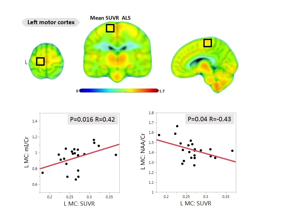
Figure 1. Top: [11C]-PBR28 SUVR maps for the ALS patients who participated in this study.
Bottom, Left: Pearson correlations between glial activation, represented by [11C]-PBR28 uptake and inflammation represented by myo-Inositol/Creatine (mI/Cr) in the left motor cortex (L MC).
Bottom Right: Pearson correlations between glial activation, represented by [11C]-PBR28 uptake and neuronal injury represented by a decline in N-acetylaspartate/Creatine (NAA/Cr) in the left motor cortex (L MC)