0616
Altered white matter microarchitecture in amyotrophic lateral sclerosis:a voxel-based meta-analysis of diffusion tensor imaging1Huaxi MR Research Center (HMRRC), Department of Radiology, West China Hospital, Sichuan University, Chengdu, People's Republic of China
Synopsis
The results of recent diffusion tensor imaging (DTI) studies on amyotrophic lateral sclerosis (ALS) have been inconclusive and controversial. We performed a voxel-based meta-analysis to identify statistical consensus between published DTI studies for altered white matter (WM) microarchitecture in ALS. Our findings provides a thorough profile of WM microarchitecture alterations in ALS and further evidence that the neuronal degeneration is not limited to the corticospinal tract but also includes the extra-motor areas, supporting the view of ALS being a multisystem degenerative disorder involving WM.
Purpose
To identify statistical consensus between published studies of diffusion tensor imaging (DTI) for altered white matter (WM) microarchitecture in amyotrophic lateral sclerosis (ALS).Methods
A systematic search was conducted for the relevant studies with voxel-wise analysis of the WM microarchitecture in ALS. Anisotropic effect-size signed differential mapping (AES-SDM) was applied to analyse the fractional anisotropy (FA) differences between ALS patients and healthy controls. Meta-regression analysis was used to explore the effects of clinical characteristics on WM integrity in patients with ALS.Results
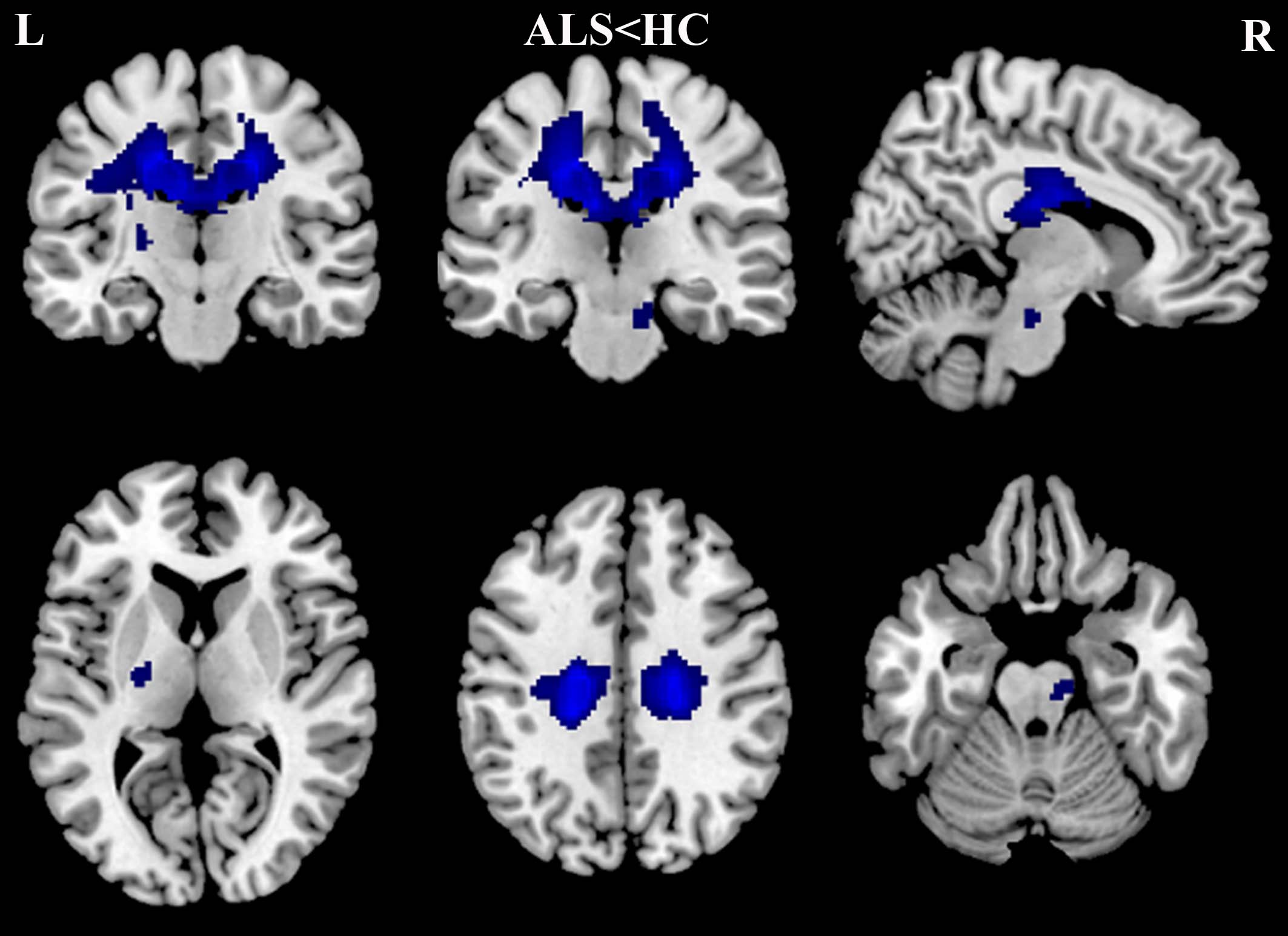
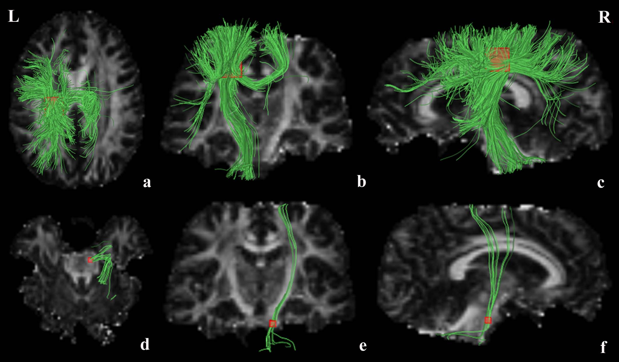
A total of 14 studies with 16 datasets including 396 patients and 360 healthy controls were identified. The pooled meta-analysis revealed that patients with ALS had significant FA reductions in two clusters relative to healthy controls, shown as in Fig. 1. The largest cluster showed a peak coordinate in the left corona radiata (MNI coordinate: x = -18, y = -26, z = 38; AES-SDM value = − 4.471, p = 0.0000; 4030 voxels) extending to the body and splenium of corpus callosum, left superior longitudinal fasciculus, posterior limb of internal capsule, right corona radiate and bilateral cingulate gyrus. Fibre tracking indicated that the main WM tracts involved in this region were the left corticospinal tract, left superior longitudinal fasciculus and the interhemispheric fibres running through the CC, shown as green tracts in Fig. 2a~c. The other cluster showed decreased FA in the right corticospinal tract (MNI coordinate: x = 8, y = -22, z = -22; AES-SDM value = − 1.839, p = 0.0031; 50 voxels) extending to right cerebral peduncle, shown as green tracts in Fig. 2d~f.
A whole-brain jack-knife sensitivity analysis of the pooled meta-analysis revealed that the decreased FA in patients with ALS in the left corona radiate was highly replicable, which was preserved throughout all 16 combinations of the datasets. The results in the right corticospinal tract remained significant in all but 4 combinations of the datasets.
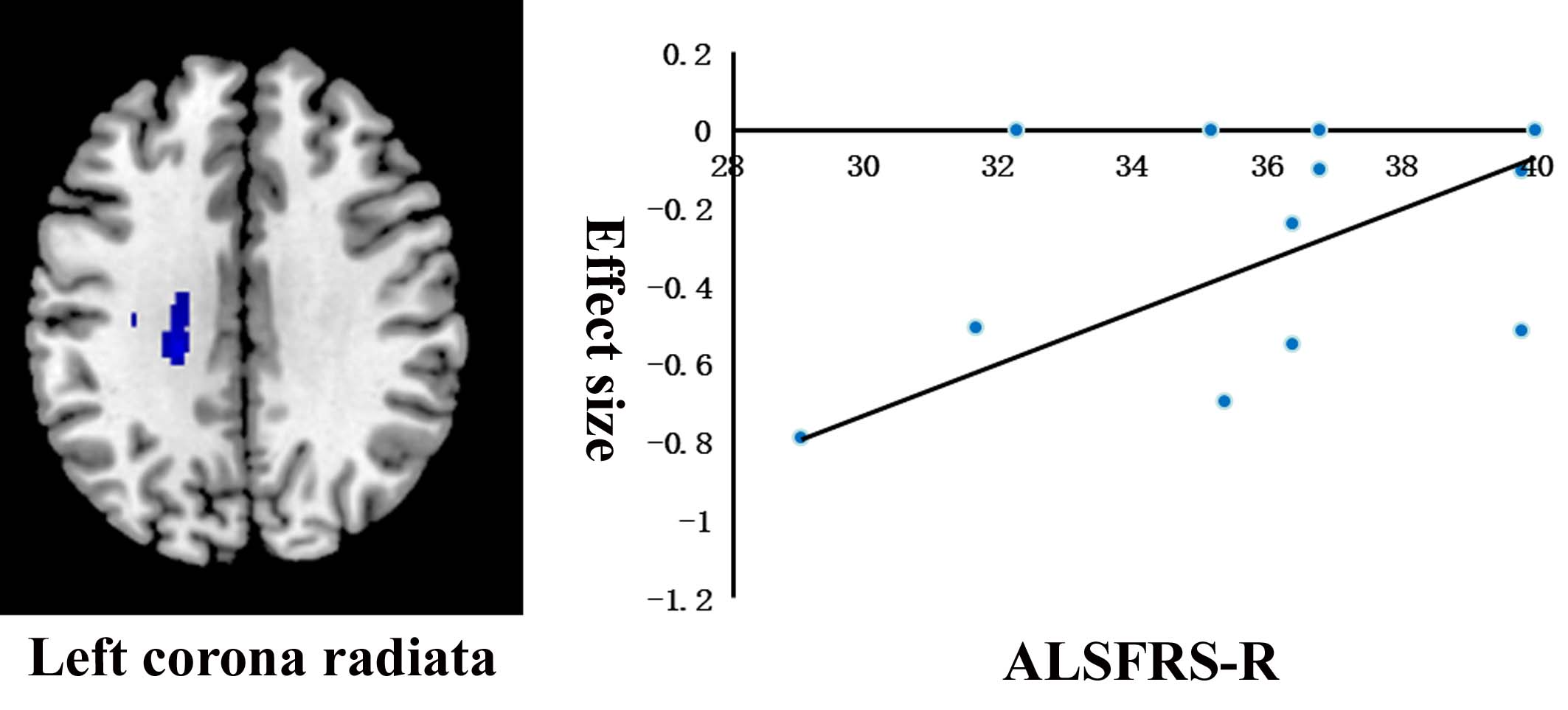
The amyotrophic lateral sclerosis functional rating scale–revised (ALSFRS-R) was positively correlated with FA reduction in the left corona radiate (MNI coordinate: x = -22, y = -28, z = 30; AES-SDM value = − 3.188, p = 0.0000; 349 voxels), shown as in Fig. 3. The mean age and illness duration were not linearly correlated with FA reductions.
Discussion
ALS is the most common adult-onset motor neuron disease that results in progressive loss of bulbar and limb function1. The etiology of ALS remains poorly understood. At present, no definitive diagnostic test or biomarker for ALS, and diagnosis considerably relies on clinical criteria2. Thus, there is an increasing necessity to identify reliable biomarkers to improve the diagnosis and objectively monitor disease progression.
Structural and functional magnetic resonance imaging (MRI) shows great potential for searching biomarkers for ALS. However, the results of recent DTI studies on ALS have been inconclusive and controversial3-6. We performed a meta-analysis for identifying the robust results of WM microarchitecture alterations to provide more insight into ALS pathogenesis. Our findings revealed an explicit impairment of the corticospinal tract connecting the precentral cortex in ALS. In addition, we also found the impairment of WM fibers connecting the extra-motor areas, including the interhemispheric fibers, the left superior longitudinal fasciculus and cingulate gyrus.
Conclusion
This study provides a thorough profile of WM microarchitecture alterations in ALS and further evidence that the neuronal degeneration is not limited to the corticospinal tract but also includes the extra-motor areas, supporting the view of ALS being a multisystem degenerative disorder involving WM.Acknowledgements
No acknowledgementsReferences
1. Zhang J, Yin X, Zhao L, et al. Regional alterations in cortical thickness and white matter integrity in amyotrophic lateral sclerosis. Journal of neurology. 2014;261(2):412-421.
2. Kiernan MC, Vucic S, Cheah BC, et al. Amyotrophic lateral sclerosis. Lancet. 2011;377(9769):942-955.
3. Agosta F, Pagani E, Rocca MA, et al. Voxel-based morphometry study of brain volumetry and diffusivity in amyotrophic lateral sclerosis patients with mild disability. Human brain mapping. 2007;28(12):1430-1438.
4. Senda J, Kato S, Kaga T, et al. Progressive and widespread brain damage in ALS: MRI voxel-based morphometry and diffusion tensor imaging study. Amyotrophic lateral sclerosis. 2011;12(1):59-69.
5. Thivard L, Pradat PF, Lehericy S, et al. Diffusion tensor imaging and voxel based morphometry study in amyotrophic lateral sclerosis: relationships with motor disability. Journal of neurology, neurosurgery, and psychiatry. 2007;78(8):889-892.
6. Keil C, Prell T, Peschel T, Hartung V, Dengler R, Grosskreutz J. Longitudinal diffusion tensor imaging in amyotrophic lateral sclerosis. BMC neuroscience. 2012;13:141.
Figures