0599
Spinal DTI Parametric Changes Following Traumatic Injury in MonkeysArabinda Mishra1, Feng Wang1, Li Min Chen1, and John C Gore1
1Radiology, Vanderbilt University Medical Center, Nashville, TN, United States
Synopsis
The proposed DTI study aims at quantification of the effect of experimentally induced dorsal column lesion at upper cervical level (C4/5) in squirrel monkeys. Diffusion parametric changes based on the directionality, and mobility of water molecules in the cellular environment, characterizes the spinal cord. Change in diffusion parameters on specific white matter tracks above and below the SCI location were compared between the lesioned and normal side of the spinal cord. A systematic group analysis of the inter-ROI changes along the white matter pathways can therefore be used to correlate the loss and recovery of sensory/motor functions over time.
Purpose
Traumatic spinal cord injury (SCI) impairs sensory, motor and autonomic functions that significantly compromises quality of patients’ life. Functional impairments are caused by a cascade of events trigged by initial insults to the spinal cord but often recover over time (weeks to months). Our previous fMRI, quantitative magnetization transfer (qMT), chemical exchange saturation transfer (CEST), and multi-parametric MRI studies [1, 2] of the SCI in monkeys have revealed complex recovery processes in both white and gray matters at and around the injury site over time. The goal of this study is to determine whether DTI (diffusion tensor imaging) measures capture white matter pathology (e.g., local demyelination) associated with targeted sections of the dorsal column white matter tract at higher cervical level, whether these changes are location (cross spinal segments) and tract specific, and these measures correlate with others (e.g., functional connectivity). We systematically quantified the diffusion parametric changes on specific white matter tracks in spinal segments above and below the injury site, and compared the measures between pre- and post-injury conditions and between normal and SCI subjects. This information provides novel insight into the potential utility of noninvasive DTI measures in monitoring and quantifying white matter pathology associated with SCI.Methods
Six squirrel monkeys were studied. Five of them underwent unilateral dorsal column lesions at higher cervical level (C4/5). All subjects were scanned using a 9.4T Agilent MRI spectrometer under isoflurane anesthesia. DTI data were acquired using a respiratory-gated, spin-echo echo-planar sequence (TR/TE=3000/30ms; voxel size= 0.5×0.5×0.5mm3), and diffusion gradients (duration/separation = 4/12ms) with b-value at 1000 s/mm2 using 30 gradient directions). High-resolution images with magnetization transfer contrast (0.25x0.25x0.5mm3, TR/TE = 220/3.24ms) were collected in the same session and used to visualize and define regions of interest (ROIs) in the DTI data (control-11 runs, lesioned-15 runs). ROIs were identified at each spinal segment (segment1-4 in Fig.1B) on both sides (normal and lesioned) within four white tracts (Fig. 1A, two lateral white matter pathways and dorsal pathway symmetrically divided into two sections). ROIs were grouped into above (rostral) and below (caudal) lesion for comparisons (Fig. 1B). The same ROI identification rule was used in normal spinal cord. An established diffusion tensor model [3] was used to calculate tensor eigenvalues. Fractional anisotropy (FA), apparent diffusion coefficient (ADC), axial and radial diffusivity (AD&RD) were measured for each ROI. We performed a pair-wise non-parametric Mann Whitney Wilcoxon (MWW) test (FWE corrected) to evaluate the statistical significance between different measures.Results
Within the dorsal column pathway (Fig. 2A), the FA values of the lesioned side (above and below) are significantly smaller than those of the normal side above lesion ROI. At the below lesion segments, the FA of the lesioned side is smaller than its normal intact side. There were no differences between above and below lesion segments in either side (lesioned or normal). Compared to the FA values of lateral white matter tracts on both sides, which are spared from injury, FA values of lesioned dorsal column (DP) tract (above and below) are significantly smaller. FA values of the dorsal column of the normal side are significantly smaller than those of lateral tracts on the same side. Within the lateral white matter tracts, FA value of the below lesion segment on the lesioned side is smaller than that of above lesion segment on the normal side (green box plots in Fig 2A). The 2-D matrix plot of significantly differed inter-ROI FA measures is shown in Figure 2B. Compared to FA values, AD and RD values are less sensitive for detecting inter-ROI differences (p-value <0.05 is considered significant for all purpose). No additional alterations were identified with AD and RD measures. ADC differences between pre- and post SCI conditions were not statistically significant across all ROIs.Discussion and conclusion
Among four DTI measures (FA, RD, AD and ADC), FA appeared to be the most sensitive indicator of the white matter tract disruption and demyelination, a finding that is consistent with our previous qMT measures. The most significant FA value reduction are observed on the lesion dorsal column tract above and below lesion site. FA value detected differences between normal and lesioned dorsal column tracts at below lesion segments. The overall FA values of dorsal column tracts of both lesioned and normal sides are significantly smaller than those of intact lateral white matter tracts. Our results demonstrated that, FA value is a sensitive measure that can differentiate white matter tract integrity and possibly demyelination state on a tract and regional (spinal segment) specific manner.Acknowledgements
This work was supported by National Institutes of Health grants R01 NS069909 (to LMC), R01 NS078680 (to JCG), and R01 NS092961 (to Gore/Chen) R01 NS093669(JCG). We would like to thank Mr. Fuxue Xin and Ms. Chaoui Tang for their helps in acquiring the MRI data and animal care.References
[1] Wang F, Qi HX, Zu Z, Arabinda M, Tang C, Gore JC, Chen LM (2015). Multiparametric MRI reveals dynamic changes in molecular signatures of injured spinal cord in monkeys. Magn. Reson. In Med., 74:1125-37. [2] Wang F, Li K, Arabinda M, Gochberg D, Chen LM, Gore JC (2016). Longitudinal assessment of spinal cord injuries in nonhuman primates with quantitative magnetization transfer. Magn. Reson. In Med., 75:1685-96. [3] Basser PJ, et al., (1996). Estimation of the effective self-diffusion tensor from NMR spin echo. Journal of Magnetic Resonance, Series B, 103(3), 247-254.Figures
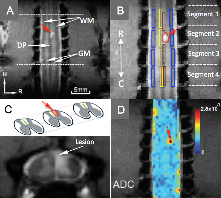
Fig.1. (A) Coronal MTC image shows gray matter (GM), lateral
white matter tracts (WM), and lesion (red arrow) on dorsal white matter pathway
(DP). (B) ROI definition: (1, 4) WM left/right (WML/WMR). H: head; R: right;
(3-4) DP left/right (DPL/DPR). Lesion is on the right side. R: rostral ROI
segment 1 above the lesion. C: caudal ROI segments 3&4 below the lesion.
(C). Axial view of the lesion, indicated by the white arrow. (D). ADC map
overlaid on anatomic (MTC) image.
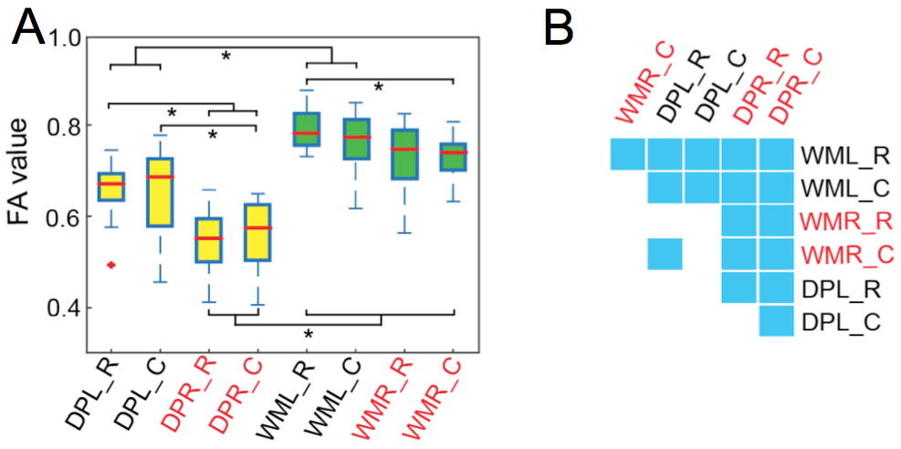
Fig. 2. (A) Box plots
of FA (fractional anisotropy) values of different ROIs. * p <0.05 (MWW test, corrected). DPL_R: left dorsal column pathway
rostral to lesion. DPL_C: left dorsal column pathway caudal to lesion. WMR_R:
lateral white matter tract rostral to lesion.
WMR_C: lateral white matter tract caudal to lesion. (B) Matrix plot of
the statistical p value that is
significant (MWW test) between two ROIs
(e.g. between WMR_C and WML_R. ROI in red font represents ROIs on the lesion
side. R and C refers to segments above and below injury location.