0590
Investigation of the role of the venous system and the glymphatic system in brain waste clearance1Department of Radiology, Wayne State University, Detroit, MI, United States, 2Neurology, Henry Ford Health System, Detroit, MI, United States
Synopsis
The recently discovered glymphatic system has become an exciting area of research because of its broad implications in both normal neurophysiological activities and neurological disorders. However, the exact relationship between the vascular system and the glymphatic system in terms of waste clearance for the brain is unclear . In addition to the glymphatic system, our preliminary MRI results suggest that the venous (but not the arterial) system also directly participates in waste removal. Fully elucidating the roles of the venous and glymphatic systems in waste removal from the brain is important for understanding the influence of waste clearance on neurological diseases.
PURPOSE
Until recently, a long-held puzzle in neuroscience had been how the brain can effectively remove its waste without a lymphatic system in the brain parenchyma. By injecting fluorescent tracers into brains of living mice, and then imaging tracer movement in real time, using two-photon microscopy, Dr. Nedergaard’s group solved the puzzle with the discovery of the glymphatic system1-4. However, due in part to technical difficulties, few studies have focused on whether the venous system, the arterial system, both, or neither also removes brain waste, as well as the relationship between the vascular and glymphatic systems. Here we present our first study of this topic with an approach that combines susceptibility weighted imaging (SWI), negative contrast agent, and blooming effects.METHODS
The multi-echo SWI scans of SD rats were performed on a 7 T magnet (ClinScan). SWI was performed for about 40 minutes once before Fe-Dextran injection and then was repeated twice to monitor and evaluate the movement of Fe-Dextran over a period of two hours. Experimental parameters: TR=60ms, TE=2.7, 7.71 ms, FA=15°, resolution of 41.6×41.6×160 µm3, intracisternal injection of FE dextran (40 kDa), and a dose of 100 μg.RESULTS
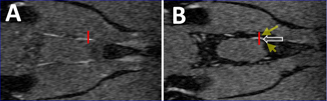
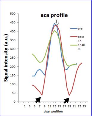
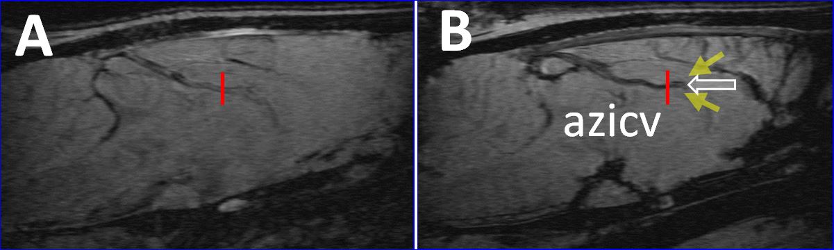
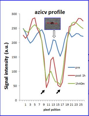
Our five key preliminary results are as follows: 1) Influx pathways are located along para-arterial spaces (dark signal due to negative contrast agent, FE-Dextran) outside of arterial vessels (bright signal due to short TE of 2.7 ms), as illustrated by comparing Figures 1A with 1B without the help of non-MRI techniques; 2) MRI tracers in the para-arterial space did not enter into the arterial blood. As illustrated in Figure 2, the intensity of the peak signal (from blood of the anterior cerebral artery, aca, labeled by an empty arrow) remained unchanged with time, while the intensities of the two valleys (the corresponding para-arterial spaces, labeled by solid black arrows) decreased first and then returned to pre-SPIO levels about 2 hours; 3) Intracisternally injected FE-dextran entered into venous blood, as illustrated in Figure 4, the peak signal intensities (from venous blood of the azygos internal cerebral vein, azicv, labeled by an empty arrow) decreased gradually with the time. These drops in signal intensities indicate the tracer entering in the vein; 4) Our proposed approach has the potential to identify all previously reported efflux pathways in one MRI study (data not shown); 5) the pathways and directions of influx/efflux systems are consistent with reports in the literature (data not shown) 1-4.DISCUSSION AND CONCLUSION
As a hydraulic system, the glymphatic system must be studied while intact, ideally with minimal invasiveness, minimal disturbance to the system’s normal function, and high sensitivity. With the exception of its low sensitivity, MRI is particularly suitable for studying the glymphatic system. Using a combination of SWI, superparamagnetic iron oxide (SPIO), and blooming effects, we have obtained results consistent with established studies of the glymphatic system 1-4. Comparing to the influx pathway, the efflux pathway is relatively less studied area. Moreover, our preliminary results suggest that the venous (but not the arterial) system is also involved in brain waste removal, potentially in concert with the glymphatic system. The results are generally consistent with differences in the anatomical structure between veins and arteries, differences in the function for waste clearance between veins and arteries outside the brain, as well as the theory that CSF exchange occurs primarily across cerebral capillaries throughout the brain parenchyma. This is the first MRI study of the glymphatic system using negative contrast agent, SWI, or their combination. Brain waste can have a broad impact on brain physiology if not removed in a timely manner. Indeed, animal studies have demonstrated the association between an impaired glymphatic system and various neurological diseases. Fully understanding the relationship between the venous and glymphatic systems in waste clearance is crucial for correctly interpreting related experimental results and for studying the role of the glymphatic system in the initiation or progression of related neurological diseases. Moreover, an MRI approach can be easily translated to human studies, a virtually unexplored area.Acknowledgements
This work was supported by NIH 1R21CA184682.References
1. Jessen NA, Munk AS, Lundgaard I, Nedergaard M. The Glymphatic System: A Beginner's Guide. Neurochemical research 2015; 40(12): 2583-2599.
2. Iliff JJ, Wang M, Liao Y, Plogg BA, Peng W, Gundersen GA, Benveniste H, Vates GE, Deane R, Goldman SA, Nagelhus EA, Nedergaard M. A paravascular pathway facilitates CSF flow through the brain parenchyma and the clearance of interstitial solutes, including amyloid beta. Sci Transl Med 2012; 4(147): 147ra111.
3. Xie L, Kang H, Xu Q, Chen MJ, Liao Y, Thiyagarajan M, O'Donnell J, Christensen DJ, Nicholson C, Iliff JJ, Takano T, Deane R, Nedergaard M. Sleep drives metabolite clearance from the adult brain. Science 2013; 342(6156): 373-377.
4. Iliff JJ, Lee H, Yu M, Feng T, Logan J, Nedergaard M, Benveniste H. Brain-wide pathway for waste clearance captured by contrast-enhanced MRI. J Clin Invest 2013; 123:1299 –1309.
Figures