0589
T2*-Weighted Echo-Planar Imaging of the Spinal Cord: Full vs. Inner Fields-of-View1Systems Neuroscience, University Medical Center Hamburg-Eppendorf, Hamburg, Germany
Synopsis
BOLD-based fMRI of the spinal cord is challenging because the high resolution required and the field inhomogeneities present promote geometric distortions in echo-planar imaging and T2*-related signal losses which both hamper the image quality and the SNR. With inner-field-of-view imaging techniques, e.g. based on spatially 2D-selective RF excitations, the acquisition can be focussed to the spinal cord which allows to minimize geometric distortions. Furthermore, shorter echo time can be achieved resulting in a significantly increased SNR that may make fMRI feasible throughout the spinal cord.
Introduction
In the past few years, interest in functional MR imaging (fMRI) of the spinal cord has risen because it performs first processing steps of sensory input and has been shown to play a major role in pain processing, even in the context of placebo analgesia (e.g. [1]). However, while fMRI with echo-planar imaging (EPI) is well-established for the human brain, spinal cord fMRI is more challenging for several reasons. First, it is located in the vicinity of the vertebral bones and the lung that both can cause significant field inhomogeneities and related artifacts, in particular geometric distortions. Second, due to the small size of the cord and its grey matter structures, a high in-plane resolution is required which increases the artifacts and the echo time and reduces the signal-to-noise ratio (SNR) accordingly. Third, for the thoracic and lumbar spinal cord, large fields-of-view (FOVs) are needed to avoid aliasing which further increases distortions and decreases the SNR. Inner-FOV imaging techniques (e.g. [2]), e.g. based on spatially 2D-selective RF (2DRF) excitations (e.g. [3]), could provide a solution to these problems as has been demonstrated for diffusion-weighted MRI (e.g. [4]). They can focus the excited magnetization to a small target regions which not only allows to minimize the FOV and related geometric distortions but also to shorten echo times and increase the SNR accordingly. In this study, T2*-weighted EPI measurements of the spinal cord obtained conventionally with full FOVs are compared to acquisitions with inner FOV based on 2DRF excitations.Methods
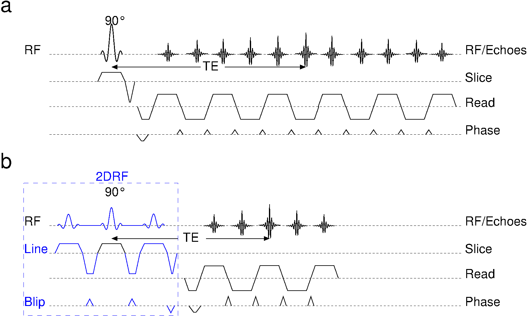
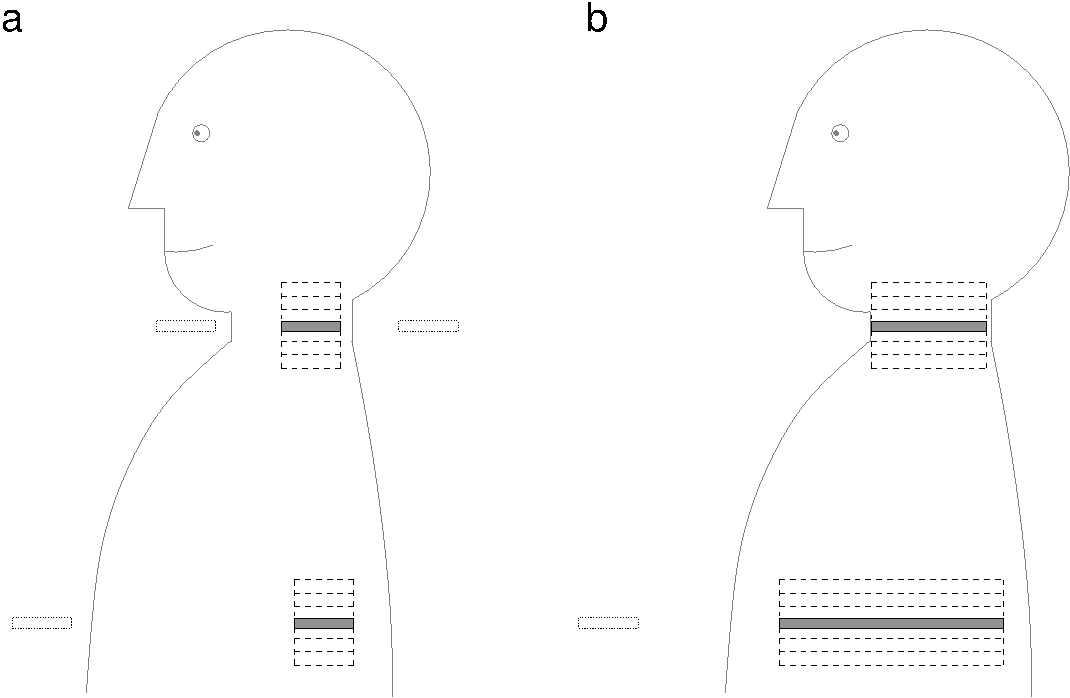
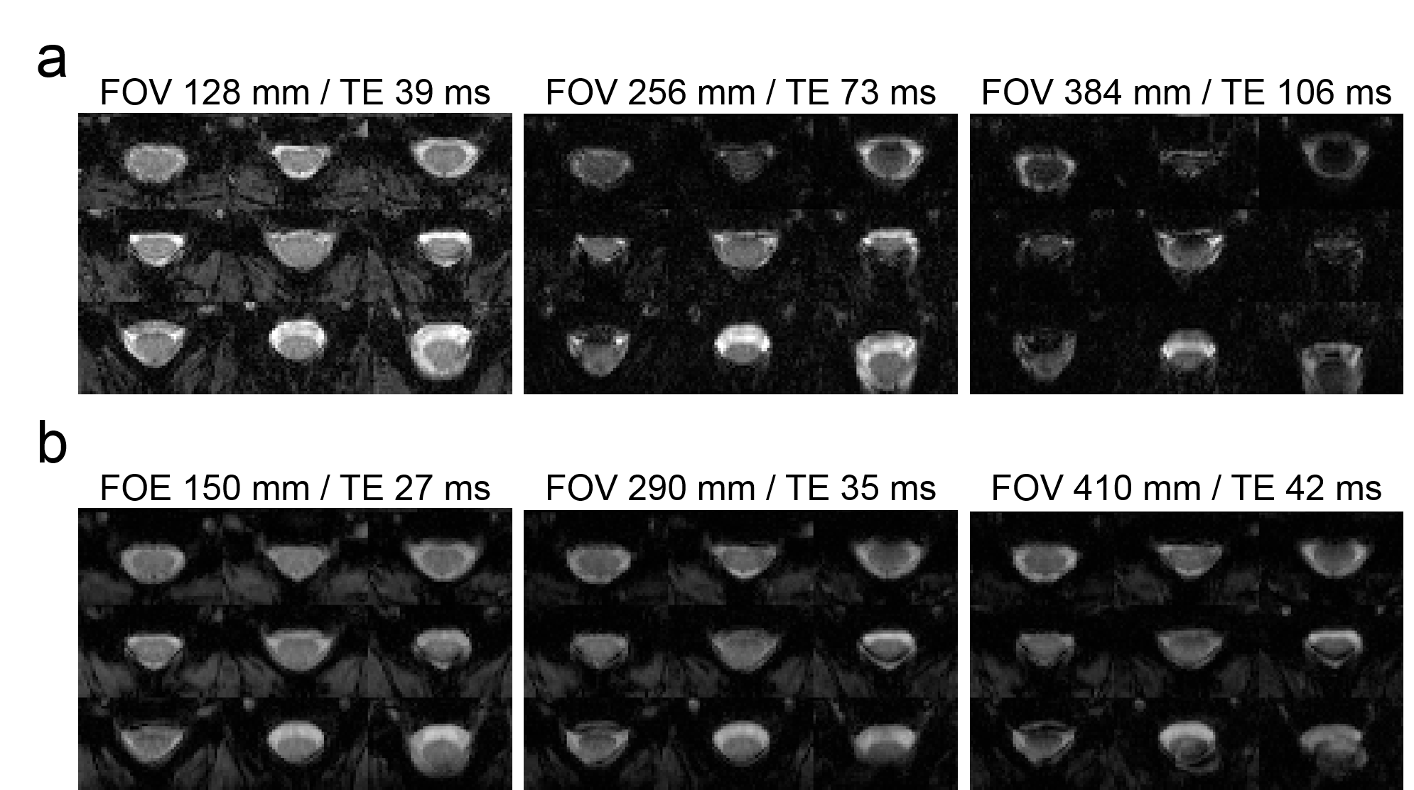
The basic pulses sequence and geometric setups are sketched in Fig. 1 and 2, respectively. For conventional EPI (Fig. 1a and 2a), the FOV in the phase-encoding must be large enough to avoid aliasing into the spinal cord. For acquisitions with 2DRF excitations (Fig. 1b and 2b), the field-of-excitation (FOE) must be large enough to position unwanted side excitations outside of the object to avoid unwanted signal contributions. Thus, both methods need to be adapted to the target spinal cord region with a larger FOV and FOE, respectively, for the thoracic and lumbar compared to the cervical spinal cord. Measurements were performed on a 3T whole-body MR system (Siemens Magnetom TIM Trio) in phantoms and in healthy volunteers from which informed consent was obtained prior to the examination. An in-plane resolution of 1.0x1.0 mm2 and a slice thickness of 5 mm was chosen as in typical spinal cord fMRI studies [1]. For all acquisitions, the echo time (TE) was minimize. Slice-selective EPI was performed with parallel acquisition techniques (acceleration factor 2) and FOVs/TEs between 128 mm / 39 ms and 384 mm / 106 ms. For inner-FOV EPI 2DRF excitation were applied with a resolution (profile sharpness) of 5.0x10.0 mm2 (slice x phase), and a fixed FOV of 32 mm (with16 mm oversampling) was used but FOEs / TEs between 190 mm / 27 ms and 410 mm / 42 ms which corresponds to very similar object sizes accessible for slice-selective and inner FOV imaging. 15 slices were covered with a slice-specific z-shim gradient pulse to minimize signal drop outs [5] and using a constant repetition time of 3500 ms that could be realized with both techniques for all parameter settings. Maps of the temporal SNR were calculated from 30 images aquired after four preparation scans.Results and Discussion
Examples images and temporal SNR maps are presented in Fig. 3 and 4, respectively. For the smallest FOV/FOE that have been chosen to be compatible with applications in the cervical spinal cord, the inner FOV techniques provides a slightly better SNR (25.4 vs. 23.8) due to the shorter echo time achievable (27 ms vs. 39 ms) and despite the reduced number of Fourier lines. The difference is more pronounced for the temporal SNR (25.5. vs. 14.0), most likely due to the sensitivity of parallel imaging techniques to motion of the spinal cord. Considering the echo times, it is obvious that it is much more beneficial, i.e. less time consuming, to increase the FOE of inner-FOV acquisitions rather than the FOV in conventional slice-selective measurements when targeting the spinal cord in larger cross sections. For the setup for the largest object size, the temporal SNR drops by less than 40% for the inner-FOV technique but by more than 60% for conventional EPI yielding values of 14.8 vs. 4.9.Conclusion
Compared to conventional slice-selective acquisitions, inner-FOV imaging techniques, e.g. based on 2DRF excitations, provide a significantly better SNR in T2*-weighted acquisitions making them possibly the method of choice for spinal cord fMRI.Acknowledgements
No acknowledgement found.References
[1] Eippert F, Finsterbusch J, Bingel U, Büchel C. Direct Evidence for Spinal Cord Involvement in Placebo Analgesia. Science 326, 404 (2009).
[2] Finsterbusch J. Functional neuroimaging of inner fields-of-view with 2D-selective RF excitations. Magn. Reson. Imaging 31, 1228-1235 (2013).
[3] Bottomley PA, Hardy CJ. Two-dimensional spatially selective spin inversion and spin-echo refocusing with a single nuclear magnetic resonance pulse. J Appl Phys 62, 4284–90 (1987).
[4] Finsterbusch J. Improving the performance of diffusion-weighted inner-field-of-view EPI based on 2D-selective RF excitations by tilting the excitation plane. J. Magn. Reson. Imaging 35, 984–992 (2012).
[5] Finsterbusch J, Eippert F, Büchel C. Single, slice-specific z-shim gradient pulses improve T2*-weighted imaging of the spinal cord. Neuroimage 59, 2307–2315 (2012) .
Figures