0585
Silent, Multi-Echo T2* Looping Star fMRI1GE Global Research, Munich, Germany, 2GE Healthcare, Orsay, France
Synopsis
In this work, we propose a novel method for quiet, 3D, T2* BOLD fMRI in form of multi-echo Looping Star. Its inaudible scanning offers unique potential for pediatric imaging, imaging auditory or speech paradigms, as well as for resting-state studies. In comparison to standard Gradient-Echo EPI, Looping Star demonstrated better tSNR and lower temporal drift. Motor activation and identification of resting state networks are demonstrated for single and combined echo processing.
Purpose
In this work, we describe a novel method for 3D, T2* BOLD fMRI, in form of multi-echo Looping Star [1]. It addresses the main limitation of low sensitivity from our previous T2 prepared ZTE method [2], while maintaining its favorable characteristics in terms of minimal geometric distortions and low/negligible acoustic noise. The method is expected to be particularly useful for pediatric imaging, auditory or speech paradigms and resting-state studies. The reduced geometrical distortions and its multi-echo capability [3] render this technique attractive for imaging regions affected by susceptibility artifacts, such as orbitofrontal cortex.Methods
Pulse sequence and reconstruction
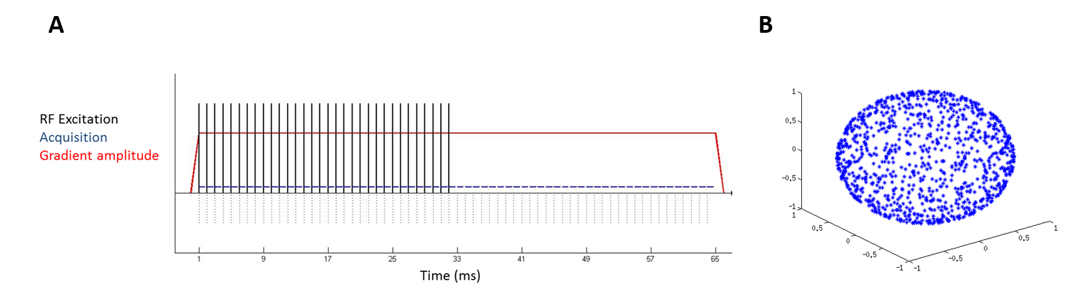
Last year, we presented Looping Star as a novel pulse sequence based on ZTE [4] and capable of acquiring multi-gradient echo data via self-refocusing. For the application to fMRI, the spatiotemporal encoding was increased (i.e. 3mm isotropic resolution, TR=2.2s, 2 echoes) by: 1) increasing the imaging bandwidth (±31.25kHz), 2) 4x sampling acceleration (w/ randomly oriented k-space loops) and 3) TGV-regularized, iterative image reconstruction.
fMRI experiment
Dual (FID+echo) and Triple-echo (FID+Echo1+Echo2) Looping Star fMRI protocols were tested using following parameters settings: flip angle = 1°, BW = ±31.25 kHz, FOV = 19.2 cm, 1024 spokes per volume, isotropic 3mm resolution, TEs= [0 26.88]/[0 13.44 26.88] ms, 32/16 spokes per loop with 840ms time per spoke, TR =2.2/3.5s.
Two healthy volunteers were scanned on a GE 3T MR750w scanner using a 12-channel head array coil (GE Healthcare, Waukesha, WI, USA). Dual-echo T2* Looping Star was run with a simple finger-tapping (FT) task (5 cycles, 100 volumes). Then, triple-echo T2* Looping Star fMRI was run with a finger-tapping task where the subject was instructed to tell to themselves the names of the finger they were touching (complex FT, 5 cycles, 100 volumes) and in resting-state fMRI with open eyes (140 volumes).
Additionally, temporal stability measurements (tSNR and temporal drift over a central area of 15x15 voxels) were performed in a silicone oil phantom (T2*~26ms) using triple-echo T2* Looping Star and a Gradient Echo EPI. Acquisition parameters were TE=26.88ms, TR=3.5s and 100 volumes for both sequences.
fMRI analyses
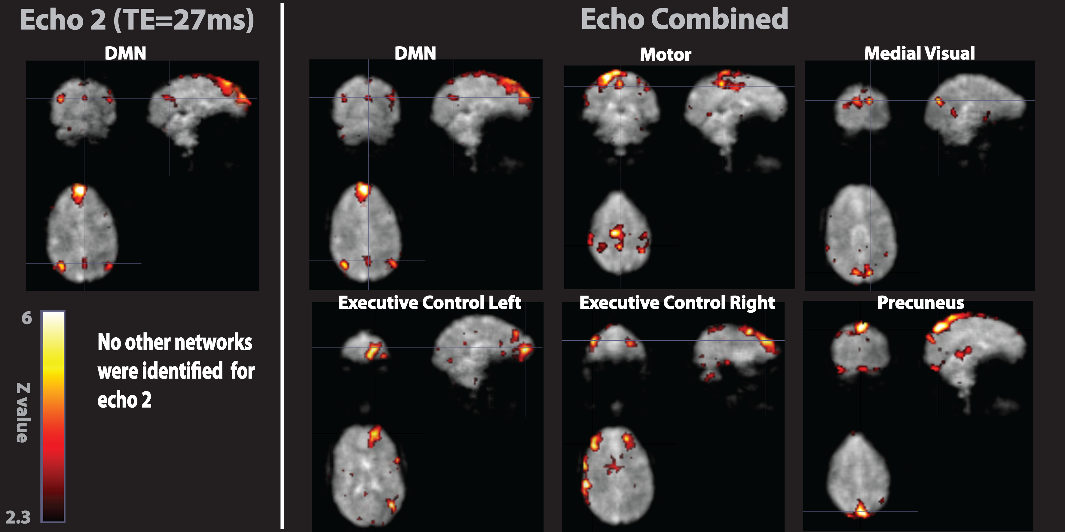
The first echo of each fMRI acquisition was used for realignment and the same transformation was applied to the other echo series. For the triple-echo fMRI, the echoes were averaged in form of a weighted sum combination [3,5]. A spatial smoothing (Gaussian FWHM 6x6x6 mm3) was applied to all the echoes series. Motor task activation statistical spatial maps were computed with a GLM analysis using an off-on boxcar function defining the paradigm model. Z score statistics were considered significant for Z>3.1, cluster-wise FWE corrected. Resting-state networks were obtained by applying Independent Component Analyses (ICA) to the preprocessed data and resting state networks were identified followed visual inspection of the independent component statistical spatial maps, time courses and frequency spectrum [6].
Results
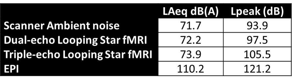
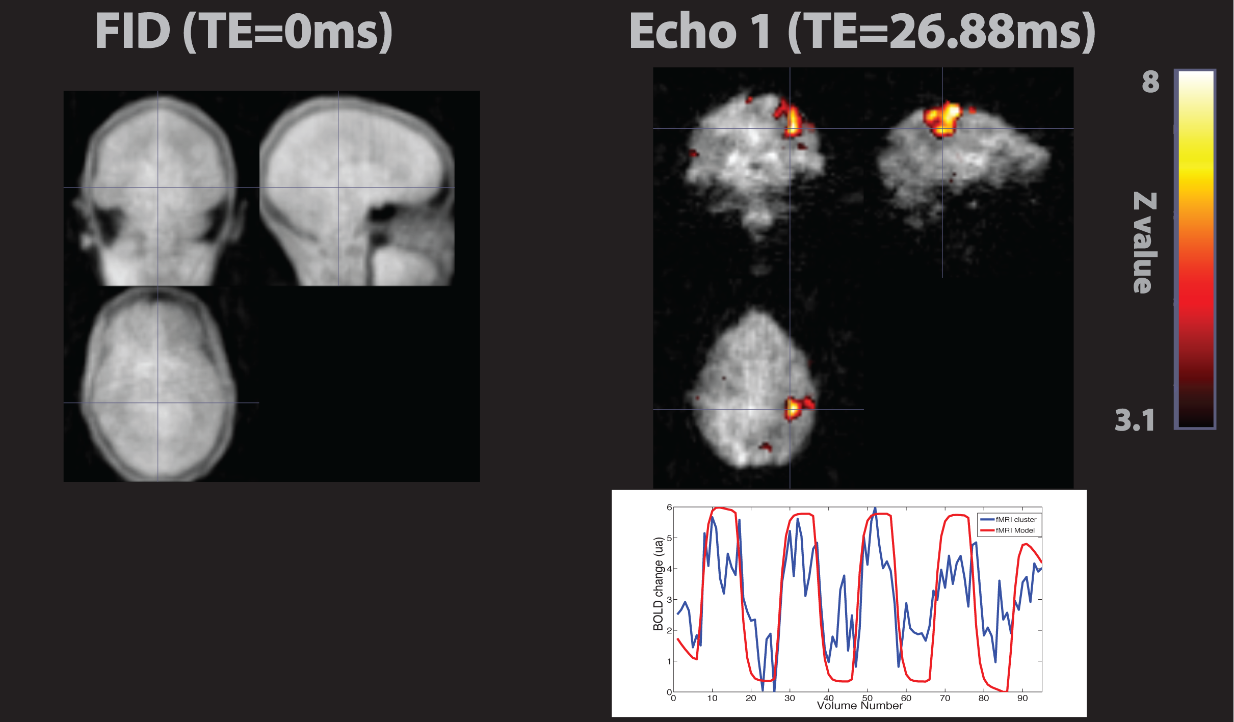
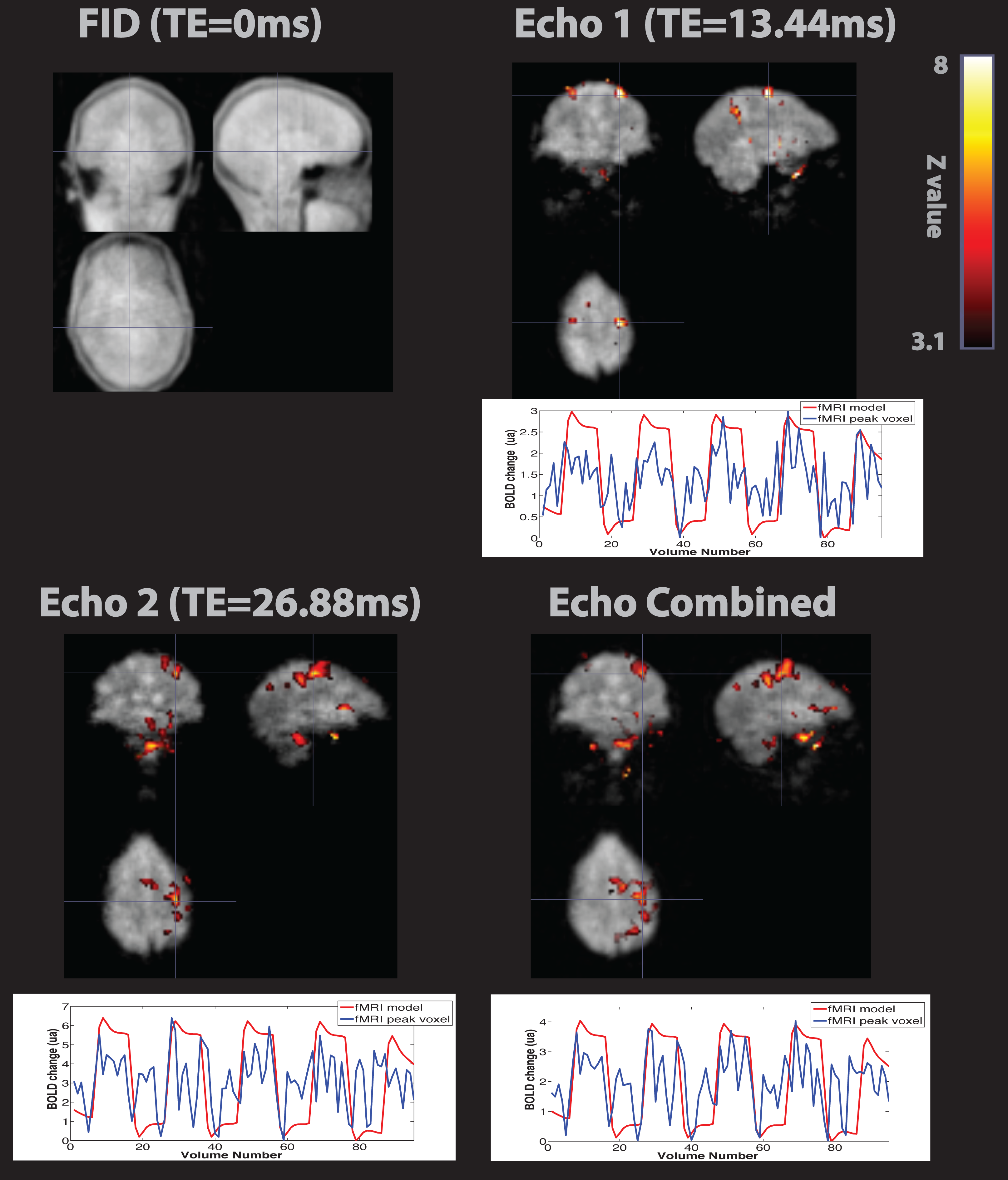
In-bore acoustic noise measurements are shown in Table 1. A temporal drift of 0.3% and tSNR=220 were obtained for second echo of the triple-echo Looping Star scan, compared to 1% drift and tSNR=150 for the Gradient Echo EPI and the same TE. Figures 2 and 3 illustrate image quality and show results from the motor tasks with dual and triple echo, demonstrating T2* BOLD contrast between 4-6% for echoes with TE=26.88ms. While the simple FT task provided activation in mainly primary (Brodmann Area- BA4) and somatosensory cortex (BA 6), the complex FT also exhibits activation in other brain areas involve in motor planning, association and memory (BA6, A20, BA40, BA24, BA 22, BA2) as expected. Figure 4 contains the results of the resting state fMRI experiment of a single subject. While only the default mode network (DMN) was identified from the echo 2 fMRI timeseries, 6 resting networks were identified from the combined echo series [7].Discussion
Here we have demonstrated quiet T2* BOLD fMRI for both task and resting-state activation using a novel, 3D multi-echo sequence called Looping Star fMRI. The sequence allows scanning with only 0.5-2dBA above the ambient noise floor of the scanner and provides full, undistorted 3D brain coverage with sufficient spatiotemporal resolution and BOLD sensitivity. This new technique also supports the benefits of multi-echo processing; like as it has been shown before for EPI2,4. Next steps include the comparison between multi-echo T2* Looping Star, multi-echo EPI and multiband multi-echo EPI in a larger population.Acknowledgements
No acknowledgement found.References
[1] U.S. Patent Application No. 15/142,957, Wiesinger F, Menini A, Solana AB entitled “SILENT MULTI-GRADIENT ECHO MAGNETIC RESONANCE IMAGING.”
[2] Solana AB, Menini A, Sacolick L, Hehn N, Wiesinger F. Quiet and distortion-free, whole-brain BOLD fMRI using T2-prepared RUFIS MR imaging, MRM Volume 75, Issue 4, 2016.
[3] Poser BA, Versluis MJ, Hoogduin JM, Norris DG. BOLD contrast sensitivity enhancement and artifact reduction with multiecho EPI: parallel-acquired inhomogeneity-desensitized fMRI, MRM 55 (6), 2006.
[4] Solana AB, Menini A, Wiesinger F. Looping star: A novel, self-refocusing zero TE imaging strategy. Singapur, ISMRM 2016
[5] Fernandez B, Leuchs L, Sämann P, Czisch M, Spoormaker V. Application of multi-echo EPI on a fear conditioning task: evidence of improved BOLD detection in ventromedial prefrontal cortex: 29 (Suppl 1):S247–S400 (338), Vienna, Austria, 2016.
[6] Kelly RE Jr, Alexopoulos GS, Wang Z, Gunning FM, Murphy CF, Morimoto SS, Kanellopoulos D, Jia Z, Lim KO, Hoptman MJ. Visual inspection of independent com-ponents: Defining a procedure for artifact removal from fMRIdata. J Neurosci Methods 189:233–245, 2010.
[7] Smith SM, Fox PT, Miller KL, Glahn DC, Fox PM, Mackay CE, Nicola Filippini N, Watkins KE, Toro R, Laird AR, Beckmann CF. Correspondence of the brain’s functional architecture during activation and rest, PNAS, vol 106, no. 31, 13045, 2009
Figures