0584
Localization of Neural Activity Using DANTE-Prepared Multi-slice EPI (DANTE-EPI) for BOLD Detection1National Institute of Mental Health, National Institute of Health, Rockville, MD, United States, 2Systems Neuroscience and Pain Lab, Stanford University, CA, United States, 3Nuffield Department of Clinical Neurosciences, FMRIB Centre, University of Oxford, Oxford, United Kingdom
Synopsis
To assess whether DANTE-EPI (Delay Alternating with Nutation for Tailored Excitation) sequence for moving blood suppressed fMRI images can effectively reduce inflow effects and, physiological noise originating from intravascular blood signal, and spurious (false positive) functional activation resulting from draining vein effects. Results were compared with images from conventional gradient echo planar imaging (GE-EPI).
Introduction:
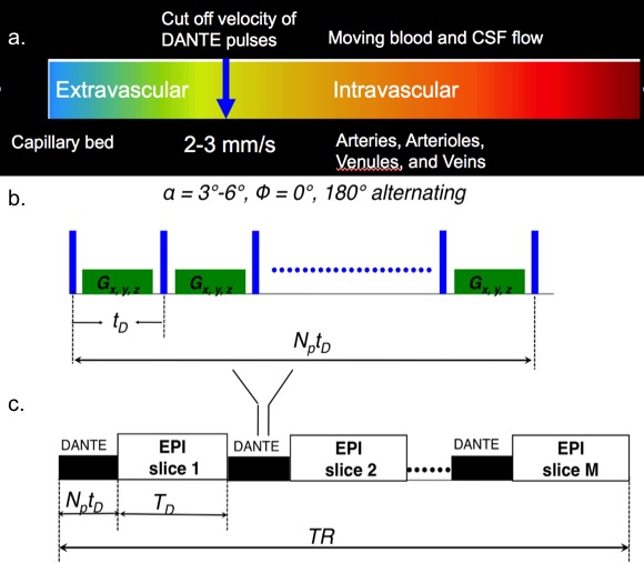
Localization and identification of neural activity detected via the blood-oxygen-level dependent (BOLD) effect with conventional GE-EPI can be severely compromised due to intravascular blood flow effects (in both arteries and veins), especially at 1.5 T and 3T. Previous studies blood flow in capillary beds of rat brain suggest that the transit flow rate of blood between vascular (arterioles and venules) and the cortical capillary bed is about 2-3 mm/sec[1]. DANTE is a novel magnetization preparation method[2] that allows selective suppression of signals from moving blood (or cerebral spinal fluid CSF) travelling with speeds greater than 2-3 mm/sec, illustrated in Fig. 1a. Previous studies suggested that DANTE preparation pulse trains in conjunction with an EPI imaging readout module should reduce contamination from large-vessel intravascular blood without significantly influencing the resulting signals derived from the parenchyma and capillary bed. In this work, we demonstrate that three major contaminants from macro-vascular blood, namely inflow effects, physiological noise, and draining vein effects, can be effectively diminished using DANTE-EPI compared with results from conventional GE-EPI, allowing improved localization of neural activity.Methods:
Ten healthy volunteers (6 males, 4 females, age 24-35 years), with written informed consent, underwent fMRI scans. DANTE-EPI sequence was illustrated in Fig. 1b and Fig.1c. All scans were acquired using a 3T Siemens Verio scanner and a 32-channel head coil. EPI parameters for all scans were: TR=2s, TE=31ms, GRAPPA=2, BW=1086 Hz/Px, FOV= 220 mm, resolution: 2.3´2.3 mm (96´96), thk=4mm, with 20% gap. Two 5 mins 19-slice series of resting state fMRI images were acquired using GE-EPI and DANTE-EPI techniques, respectively. Temporal standard deviation (tSTD) maps were calculated from the resting state data, and overlaid on high-resolution T1 images for comparisons. An additional 5.5 mins 11-slice task-related protocol was carried out, using a right-hand finger-tapping task (1 Hz) with 30s ON-OFF blocks. DANTE parameters for the resting-state acquisition were: number of pulses (Np) 44 for each DANTE preparion duration 22 ms; inter-pulse delay (td) 0.5-0.7 ms; flip angle (FA) 6°; for the task-based sequence FA=3° and Np=212 was used for each DANTE preparion duration 106 ms.Results:
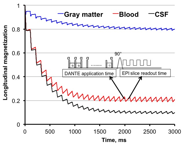
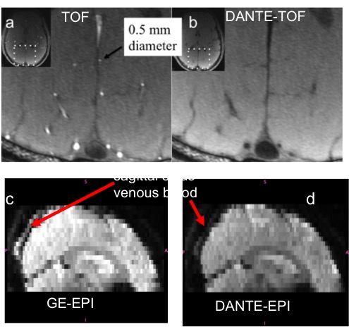
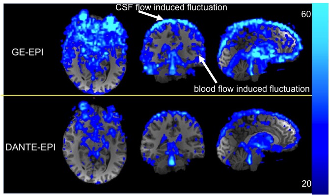
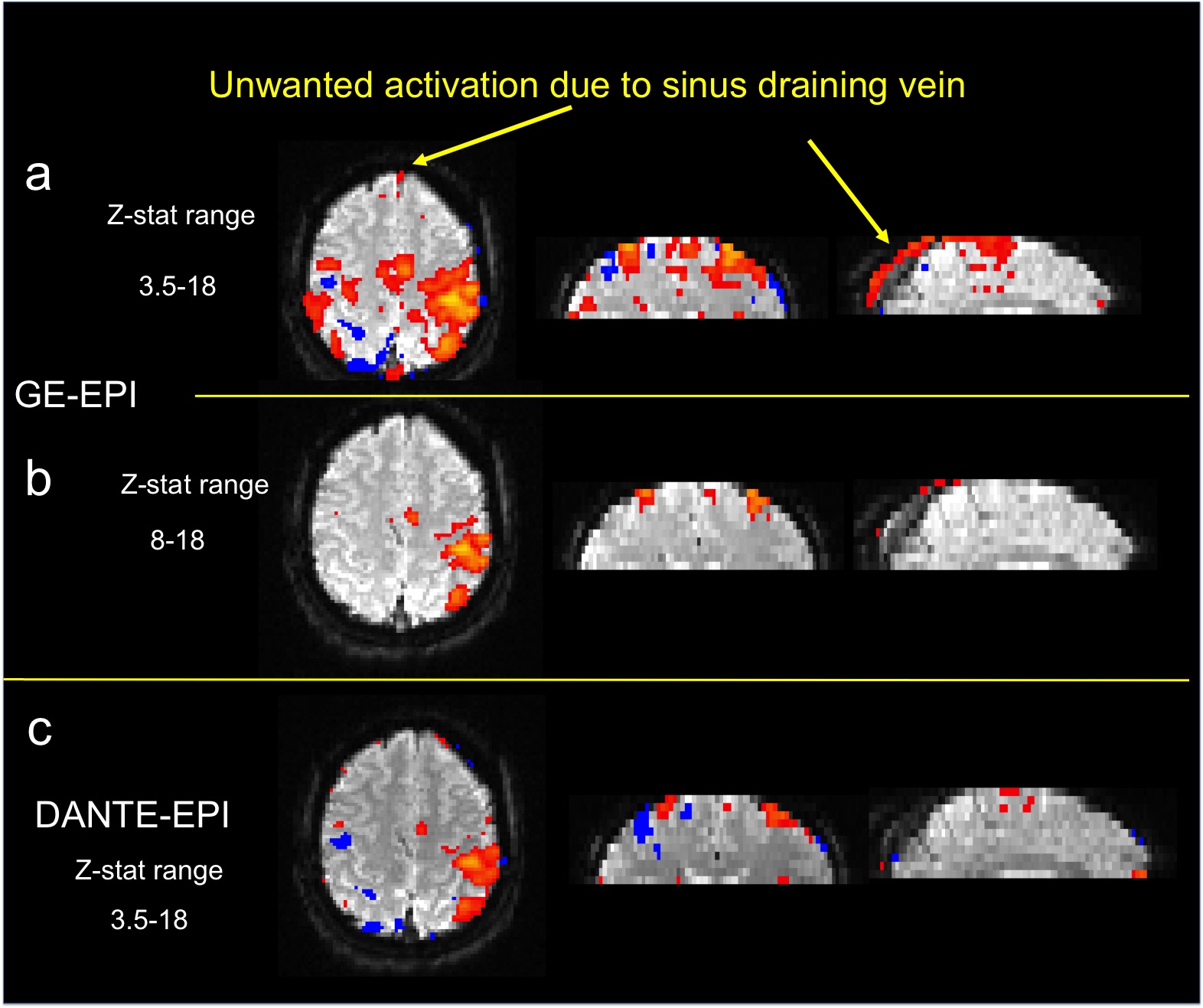
Bloch simulations of the 19-slice protocol are shown in Fig. 2, demonstrating the transient longitudinal magnetization evolution of moving blood, CSF and static gray matter tissue at the start of the DANTE preparation. 1. Elimination of in-flow effect: To demonstrate mitigation of in-flow effects when DANTE pulses are applied, high resolution (0.3´0.3mm) time-of-flight (TOF) data were acquired, as shown in Fig. 3a, showing hyper-intense arterial signals due to a strong in-flow effect, which is greatly reduced with DANTE preparation, as shown in Fig. 3b. In addition, sagittal images reconstructed from multi-slice GE-EPI and DANTE-EPI are shown in Figs. 3c and 3d. It can be visually observed that blood signal in the sagittal sinus is largely suppressed in DANTE-EPI image. 2. Temporal standard deviation maps calculated from the GE-EPI and DANTE-EPI resting state data, and arising mainly from cardio-respiratory physiological motion of CSF and blood, are shown in Fig. 4. The relative color scale represents the detected physiological noise. Significant signal suppression of moving blood and CSF physiological fluctuations is observed in the images from the DANTE-prepared EPI sequence. 3. Removal of draining vein effects: The extent of spurious activations in the sagittal sinus vein, shown in Fig. 5a from GE-EPI, is substantially diminished in images from the DANTE-EPI scans, in Fig. 5c, suggesting considerable improvement in identifying the true activation region. In terms of BOLD sensitivity, it shows that activation regions from DANTE-EPI data, in Fig. 5c appear to have comparable sensitivity when compared with Fig.5b, which was the same data with Fig. 5a but with increased minimal z-stat for filtering out the fake positive activation at sinus vein.Conclusion:
Detection of relevant cortical activation with DANTE-EPI techniques appears to be more closely localized to the microvasculature and the associated tissue bed, compared with the GE-EPI technique. Therefore, adoption of DANTE-EPI may be helpful in improving the localization of neural activity in fMRI studies.Acknowledgements
This work was supported by the British Heart Foundation, UK, the Dunhill Medical Trust.References
1. Ivanov, K. P., M. K. Kalinina, and Yu I. Levkovich. "Blood flow velocity in capillaries of brain and muscles and its physiological significance." Microvascular research 22, no. 2 (1981): 143-155.
2. Li, Linqing, Karla L. Miller, and Peter Jezzard. "DANTE prepared pulse trains: A novel approach to motion sensitized and motion suppressed quantitative magnetic resonance imaging." Magnetic resonance in medicine 68, no. 5 (2012): 1423-1438.
Figures