0572
Mitigation of bSSFP Flow Artifacts using Partial Dephasing1Electrical Engineering, Stanford University, Stanford, CA, United States, 2Radiology, Stanford University, Stanford, CA, United States
Synopsis
We show that partial dephasing mitigates artifacts from through-plane flow near dark bands in balanced SSFP. A 30o-60o range in the phase accrual during a TR is created over the voxel by slightly unbalancing the slice-select rephaser. The effects of partial dephasing on the spectral profiles for various flow rates were simulated, and the simulations were validated in a flow phantom. By decreasing the strength and non-linearity of the signal’s dependence on through-plane flow rate, partial dephasing mitigates the transient artifacts caused by pulsatile flow. In volunteer studies, it noticeably decreased artifacts in all of the phase-cycled cardiac cine datasets acquired.
Purpose
Balanced steady-state free precession (bSSFP) is commonly used for cardiac imaging due to its high SNR and desirable myocardium-blood contrast but is sensitive to off-resonance. In slice-selective acquisitions, through-plane flow near a steady-state stop-band (or "near-band") can cause strong enhancement from out-of-slice spins and artifacts propagating in the phase-encode direction from the band.1-3 In addition, these artifacts make multiple-acquisition bSSFP, which is normally used to get banding-free bSSFP images, challenging in the heart since the band falls in the blood pool in at least one of the phase-cycled component images.4-6 Here, we propose using partial dephasing7 to mitigate near-band flow artifacts, preventing them from corrupting other parts of the image and aiding multiple-acquisition bSSFP in the heart.Methods
In partial dephasing, a 30o to 60o range in the phase accrual during a TR is created over the voxel by slightly unbalancing the slice-select refocusing lobe before the RF pulse.7 In standard bSSFP, at the bands, spins that flow out of the slice remain in-phase, causing a manyfold increase in the signal.1 We hypothesized that partial dephasing would decrease this out-of-slice coherency by causing spins that have flown out to experience a different amount of phase accrual each TR. By therefore decreasing the strength and non-linearity of the dependence of the signal at the bands on the through-plane flow rate, partial dephasing would mitigate the transient artifacts caused by pulsatile flow.
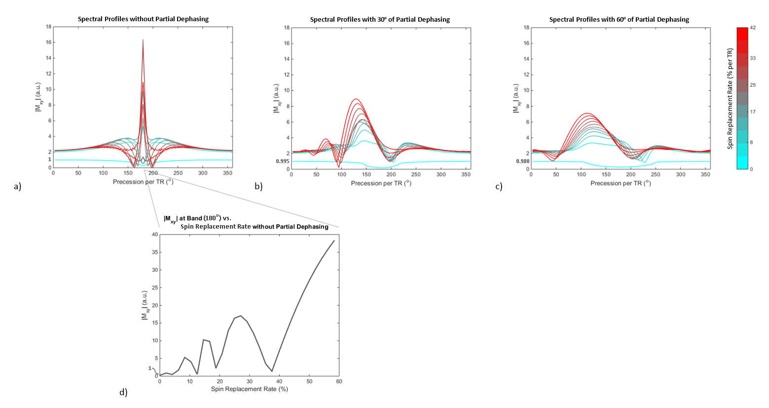
To test this, the spectral profiles for various flow rates with a fully-balanced sequence and with 30o and 60o of partial dephasing were determined using Bloch simulations similar to those used by Markl and Pelc.1 The parameters were: TR=3.5 ms, TE=1.75 ms, T1=1000 ms, T2=150 ms (in-slice), and T2*=75 ms (out-of-slice). Spins were moved through the slice profile of a 60o TBW-2 Hamming-windowed sinc with FWHM of 6 mm. Static spins and flows up to 100 cm/s, corresponding to 58.3% spin replacement per TR, were simulated. The level of inconsistency of the spectral profile across different flow rates was used as an indication of how much variable flow during image acquisition will cause the signal to vary, thus leading to transient-related artifacts.
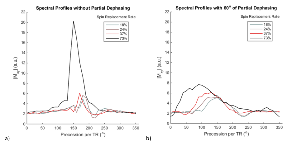
To validate the simulations, we imaged a slice through a constant flow phantom with and without 60o of partial dephasing with 36 different RF phase-cycling amounts. Imaging was done at 1.5 T with 4.6 ms TR and 2.0 ms TE. Various slice thicknesses (8, 6, 4, and 2 mm) resulted in different spin replacement percentages. To get empirical spectral profiles, the mean signal in an ROI in the tube, normalized by slice thickness, was plotted versus phase-cycling amount. The average velocity in the ROI ranged from 28.8 to 35.1 cm/s during the scan session.
Cardiac cine images of axial slices of six volunteers were acquired with 0o, 30o, and 60o of partial dephasing to assess if partial dephasing mitigates near-band flow artifacts. Images with 180o and 0o phase-cycling were acquired with 3.4 ms TR, 1.4 ms TE, and 6-8 mm slice thickness. The Y linear gradients were offset from the optimal shim to create increased off-resonance.
Results
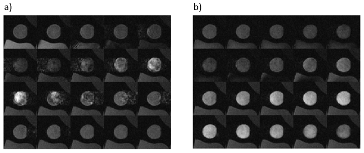
Bloch simulations indicate that the strength and non-linearity of the dependence of the near-band signal on the through-plane flow rate in fully-balanced SSFP is substantially reduced by partial dephasing – with 30o and especially 60o of partial dephasing, the spectral profiles in Figure 1 morph smoothly as a function of the flow rate. Therefore, simulations suggest that partial dephasing may mitigate near-band flow artifacts while maintaining bSSFP’s high SNR. The spectral profiles measured in the flow phantom (Figure 2) validate the simulations. With partial dephasing, the images of the lumen at frequencies surrounding the band do not have the flow artifacts seen without partial dephasing (Figure 3).
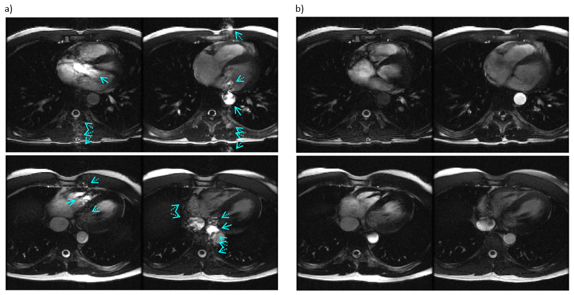
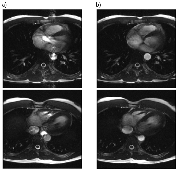
In vivo, partial dephasing removes the artifacts seen in standard bSSFP images (Figure 4). The root-sum-of-squares-combined8 multiple-acquisition images with partial dephasing do not have obvious artifacts, in stark contrast with the images without partial dephasing (Figure 5).
Discussion and Conclusion
Flow phantom and in vivo images indicate that partial dephasing alleviates the near-band flow-related artifacts to which bSSFP is sensitive. In our experience, artifact mitigation was robust, with a marked improvement in several cardiac phases of all of the cine datasets acquired. Sixty degrees of partial dephasing suppressed artifacts slightly more robustly than 30o but may broaden stop-bands, so the optimal amount should be investigated.
Although it does not address the signal nulls, partial dephasing prevents flow artifacts from dark bands from corrupting other parts of the image. It therefore facilitates multiple-acquisition bSSFP in the heart, which is banding-free.
Acknowledgements
Thank you to the Fannie and John Hertz Foundation, NSF Graduate Research Fellowship Program, and GE Healthcare for their support. In addition, we would like to thank Brian Hargreaves for a helpful discussion.References
1. Markl M, Pelc NJ. On flow effects in balanced steady-state free precession imaging: pictorial description, parameter dependence, and clinical implications. J Magn Reson Imag 2004;20:697-705.
2. Saremi F, Grizzard JD, Kim RJ. Optimizing cardiac MR imaging: practical remedies for artifacts. RadioGraphics 2008;28(4):1161-1187.
3. Schar M, Kozerke S, Fischer SE, Boesiger P. Cardiac SSFP imaging at 3 Tesla. Magn Reson Med 2004;51:799-806.
4. Wang Y, Shao X, Martin T, Moeller S, Yacoub E, Wang DJ. Phase-cycled simultaneous multislice balanced SSFP imaging with CAIPIRINHA for efficient banding reduction. Magn Reson Med 2015. doi: 10.1002/mrm.26076.
5. Datta A, Baron CA, Ingle RR, Cheng JY, Nishimura DG. Breath-held phase-cycled cardiac cine MRI using slow frequency modulation. In Proceedings of the 24th Annual Meeting of ISMRM, Singapore, 2016. 3214.
6. Fischer A, Hoff MN, Ghedin P, Brau ACS. Banding-artifact free bSSFP cine imaging using a Geometric Solution approach. In Proceedings of the 24th Annual Meeting of ISMRM, Singapore, 2016. 1827.
7. Epstein FH, Kim D, McVeigh E. Reducing signal oscillations during the approach to steady state in true FISP using partial dephasing. In Proceedings of the 9th Annual Meeting of ISMRM, Glasgow, Scotland, UK, 2001. 1786.
8. Bangerter NK, Hargreaves BA, Vasanawala SS, Pauly JM, Gold GE, Nishimura DG. Analysis of multiple-acquisition SSFP. Magn Reson Med 2004;51:1038–1047.
Figures