0553
As Easy as Echo: Interactive Fetal Cardiac MR Imaging1Advanced Clinical Imaging Technology, Siemens Healthcare AG, Lausanne, Switzerland, 2Department of Radiology, University Hospital (CHUV) and University of Lausanne (UNIL), Lausanne, Switzerland, 3LTS5, École Polytechnique Fédérale de Lausanne (EPFL), Lausanne, Switzerland, 4Center for Biomedical Imaging (CIBM), Lausanne, Switzerland, 5Department of Pediatrics, University Hospital (CHUV) and University of Lausanne (UNIL), Lausanne, Switzerland, 6Department of Gynecology-Obstetrics, University Hospital (CHUV) and University of Lausanne (UNIL), Lausanne, Switzerland, 7Magnetic Resonance, Siemens Healthcare GmbH, Erlangen, Germany
Synopsis
Although fetal echocardiography remains the gold standard for prenatal detection of congenital heart disease, mainly due to its ease-of-use, availability, and high diagnostic performance, MRI is occasionally used as a complementary and safe modality. However, MRI workflow is problematic as bulk fetal motion can occur anytime during acquisition, but can only be identified retrospectively, after image reconstruction. We describe an acquisition scheme that changes this workflow and allows interactive real-time planning of the fetal cardiac scan. The operator can easily find the desired scan plane even in a moving imaging target. This technique is applied and tested in two pregnant patients.
Purpose
Congenital heart disease (CHD) occurs in about 9/1000 live births and is a leading cause of infant mortality. Prenatal detection of CHD allows for informed decision-making, early referral to specialized care centers, and can improve the final outcome. Fetal echocardiography is currently the clinical modality of choice for its safety, easy accessibility, low cost, real-time imaging capabilities and high sensitivity for detecting major CHD1. However, fetal echocardiography is highly user-dependent, sometimes limited by acoustic windows, difficulties in imaging the distal vasculature, fetal position, maternal obesity or abdominal wall scarring form prior surgeries. These limiting factors can potentially be overcome by MRI2. However, unpredictable bulk fetal movements – which are usually a minor problem for echocardiography – complicate the MRI acquisition as fetal motion can only be identified retrospectively, after image reconstruction.
We propose an acquisition and reconstruction scheme that provides on-line real-time information and allows interactive planning of the fetal cardiac scan, with which – similarly to echocardiography – the operator can easily navigate the scan plane to the anatomy of interest in real-time, even if fetal bulk motion occurs. More detailed information about cardiac anatomy and function can then be extracted from the same data off-line, after the scan.
Methods
Data Acquisition: Data acquisition is performed with a prototype, non-triggered, 2D radial golden-angle bSSFP trajectory3. The trajectory is constructed such that all angles are repeated every N k-space lines. As the close form of the golden ratio is proportional to the limit of the ratio between two subsequent numbers of the Fibonacci series, by choosing N as a high Fibonacci number, the repeated series becomes the close approximation of a continuous golden-angle acquisition (e.g. for N=6765, the (N+1)th angle will be reset to 0° instead of 0.0119°). In such repeated acquisition, regridding parameters can be pre-calculated for fast real-time on-line reconstruction. Moreover, as acquisition and reconstruction can be interconnected through a real-time feedback-loop4, the user can interactively modify position and orientation of the imaging slice while scanning (Fig.1). The temporal resolution of the interactive real-time acquisition was optimized in adult subjects. The interactive feature allows assessment of the slice position and orientation changes at the scanner in real-time, similar to moving an echocardiography probe (Fig.2). Once the desired fetal cardiac view is found, the operator can monitor fetal movements during the maternal breath-hold and respond with scan plane adjustments as needed. The last few seconds of image data acquired during a period that is free of fetal bulk motion are then also transferred for off-line compressed sensing reconstruction with a much higher temporal resolution.
Offline Image Reconstruction: As the above workflow ensures that the last part of the acquisition was performed during a maternal breath-hold and without fetal bulk motion, data are also reconstructed off-line using a previously published k-t sparse SENSE algorithm5. This results in images of the fetal heart with high quality and high temporal resolution. Briefly, the reconstruction process consists of three steps: (1) k-t sparse SENSE reconstruction of all motion-free data acquired during the breath-hold, (2) extraction of a self-gating cardiac signal, and (3) self-retro-gated reconstruction of one single heartbeat (50% shared phases).
Patients: Datasets from two patients (gestational age: 32 and 31 weeks) were acquired and reconstructed with the proposed methodology. Parameters: 1.5T clinical scanner (MAGNETOM Aera, Siemens Healthcare), TE/TR=1.99/4.1ms, field-of-view=(260x260)mm2, matrix size=(256x256)pixel, pixel size=(1.0x1.0x4.0)mm3, RF-excitation angle=70°, bandwidth=1028Hz/pixel. Results were visually compared with those obtained with a previously described non-interactive acquisition methodology6.
Results
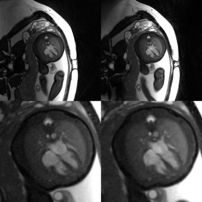
Temporal resolution for the interactive planning was 0.5s per frame. This was sufficient to navigate to the anatomy of interest in real-time, to make adjustments in response to fetal bulk motion, and to monitor the maternal breath-hold (Fig.2). Acquisitions in both patients were successful. All steps of the off-line reconstruction described above could successfully be completed (Fig.3 and Fig.4). The temporal resolution after off-line reconstruction was 12.5ms. The datasets acquired with the interactive technique achieved the same image quality as those reconstructed from the non-interactive acquisitions6. Ventricles, atria, great vessels, papillary muscles and valves, could easily be visually identified (Fig.5).Discussion and Conclusion
A new technique which combines the ease-of-use of interactive real-time acquisitions and advanced off-line reconstruction at high temporal resolution allows an easy planning of dynamic fetal cardiac MRI scans as well as a high quality imaging output. The interactive features of this technique at the scanner are similar to those of echocardiography, as the operator can freely move and rotate the imaging plane, while relying on a real-time visual feedback. Fetal movements and maternal breath-holds could be successfully monitored in all patients.Acknowledgements
This work was supported by the Swiss National Science Foundation grants 320030_143923 and 326030_150828References
1. Votino C, et al. Magnetic resonance imaging in the normal fetal heart and in congenital heart disease. Ultrasound Obst Gyn. 2012;39(3):322-329.
2. Wielandner A, et al. Potential of magnetic resonance for imaging the fetal heart. Seminars in Fetal Neonatal Medicine. 2013;18:286-297.
3. Winkelmann S, et al. An Optimal Radial Profile Order Based on the Golden Ratio for Time-Resolved MRI. IEEE Trans on Med Imaging. 2007;26(1):68-76.
4. McVeigh ER, et al. Real-time, interactive MRI for cardiovascular interventions. Acad Radiol. 2005;12(9):1121-1127.
5. Yerly J, et al. Coronary endothelial function
assessment using self-gated cardiac cine MRI and k-t sparse SENSE. Magn Reson
Med. 2016; Early view
6. Chaptinel J, et al. A Golden-Angle Acquisition Coupled with K-T Sparse SENSE Reconstruction for Fetal Self Retro-Gated Cine Cardiac MRI: An in Vivo Feasibility Study. ISMRM. 2016;24:459.
Figures
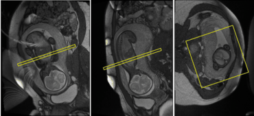
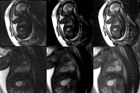
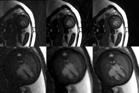
Figure 3: Offline reconstruction of 2-chamber view. The last part of the interactive real-time acquisition (during
maternal breath-hold and free from bulk fetal motion) is reconstructed off-line with a methodology previously described5. The
figure shows - from left to right - the last part of the interactive real-time acquisition, the first step
of the off-line reconstruction, and the final retro-self-gated reconstruction at
high temporal resolution. The full dataset is shown in the upper row, while the
zoomed image of the heart is displayed in the lower panel. Notice the differences in temporal resolution (0.5s / 12.5ms / 12.5ms) and image quality.