0543
Integrated T2 preparation and Inversion Recovery pulse (T2IR) for combined myocardium T1 and T2 mapping1Center for Biomedical Imaging Research, Department of Biomedical Engineering, School of Medicine, Tsinghua University, Beijing, People's Republic of China
Synopsis
In this study we designed a novel hybrid T1 and T2 magnetization preparation pulse (T2IR) and implement a combined T1 and T2 mapping sequence based on MOLLI scheme. Phantom experiments showed that the proposed sequence has high consistency with reference methods for both T1 (R2= 0.99) and T2 (R2=0.97) measurements. In vivo results showed that high quality T1 and T2 maps of myocardium could be obtained by the proposed sequence.
Background
In cardiac magnetic resonance, T1 and T2 maps are used as non-invasive tools for quantitatively1 myocardial tissue characterization. Simultaneous measuring T1 and T2 maps in a single scan with the intrinsic advantages of high scan efficiency and inherent co-registration between maps. Several techniques have been explored, such as MR fingerprinting2 and Joint T1-T23, though the map quality is not that ideal. MOLLI4 has been wildly used for T1 mapping because of its robustness and high image quality.
In this study,
we sought to develop a combined T1 and T2 mapping sequence borrowed the MOLLI
acquisition scheme by using an integrated T2 preparation and inversion recovery
pulse (T2IR) dedicated designed for a hybrid T1 and T2 magnetization
preparation.
Methods
All imaging experiments were performed on a 3T MR scanner (Achieva TX, Philips Healthcare, Best, Netherlands). An 8-channel head coil was used for the phantom experiments, and a 32-channel cardiac coil for the in vivo experiments.
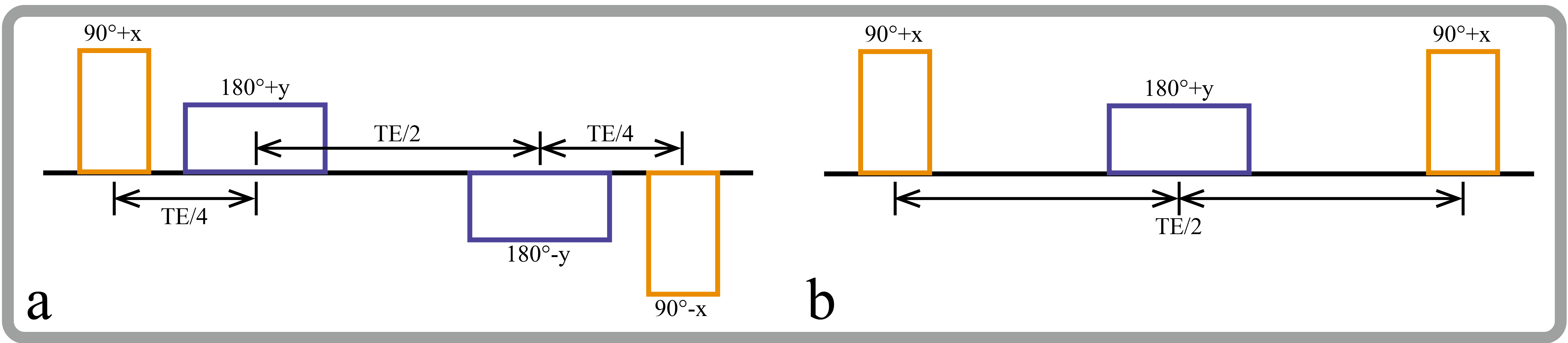
T2IR:
A typical conventional T2 preparation pulse5 is shown in Figure 1a. It is composited by two 90° RF pulses (90°+x, 90°-x) and a pair of refocusing pulses (180°+y, 180°-y) in between the two 90° pulses. We constructed the T2IR pulse by changing the phase of the second 90° RF pulse to +x and removing one refocusing pulse (Figure 1b). T2IR pulse is sort of simultaneous T2 preparation and inversion which can provided hybrid T1 and T2 weighting.
Sequence design:
The proposed combined T1 and T2 mapping sequence, which was dubbed as T2IR-Molli, employed the same acquisition scheme as the original Molli(3-3-5) sequence except for the replacement of the inversion pulse by the proposed T2IR pulse. T2 preparation echo times of T2IR pulse and the inversion recovery times were 10/130ms, 25/130ms, 45/130ms for the three Look-Lock acquisition units, respectively. Pixel-wise fitting of T1 and T2 map were based on the following model:
S= A-(A+B*exp(-TE/T2))*exp(-TD/T1*))
T1 = B/A*T1*
where TE and TD are echo time of T2 preparation and inversion recovery time in T2IR, respectively. S is the original signal intensity, A and B are the fitting model coefficients, T1* is the apparent T1.
Phantom experimen:
Fifty phantom vials with different T1 (500-2000ms) and T2 (30-80ms) values were made and underwent a comparison scan among the proposed sequence, an inversion recovery spin-echo scan for reference T1 measurement, and a CPMG spin-echo scan for reference T2 measurement.
In vivo experiments:
Approved by the local institutional review board, 5 healthy volunteers (4 males, mean age=25) without known cardiovascular disease were recruited for in-vivo study with written informed consent. T2IR-Molli, Molli, and the conventional 4 Images T2 mapping (4IMGST2)6 were performed with breath holding manner.
Shared acquisition parameters for all the three scans were: FA=35°, SENSE factor=2, partial echo=0.85, voxel size=1.7x2.1x8mm3, and linear profile order for bSSFP readout.
Results
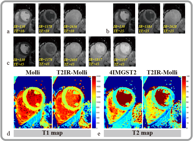
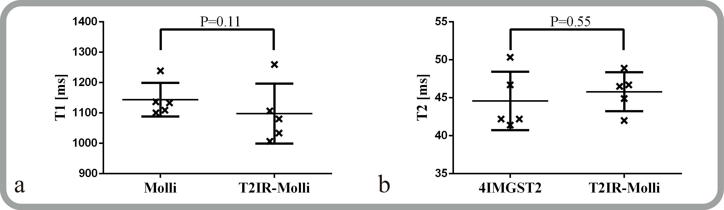
In phantom experiment, T2IR-Molli has excellent correlations with the reference methods for both T1 (R2=0.99, p<0.001, N=15) and T2 (R2=0.97, p<0.001, N=15) measurement. Representative images of a healthy volunteer from T2IR-Molli were shown in Figure 2a, 2b and 2c corresponding to the three Look-Lock acquisition units. The T1 and T2 maps from T2IR-Molli, Molli and 4IMGST2 were shown in Figure 2d and 2e. Mean myocardium T1 values of all subjects measured by MOLLI and T2IR-Molli were 1144 ± 55.3 ms and 1098 ± 98.6 ms, respectively (Figure 3a). Mean myocardium T2 values measured by 4IMGST2 and T2IR-Molli were 44.6 ± 3.8 and 45.8 ± 2.5 ms, respectively (Figure 3b).
Conclusion
In this study, we proposed a novel preparation pulse T2IR that allows hybrid T2 and T1 magnetization preparation. Combined with the original Molli sequence, T2IR enables combined T1 and T2 mapping with high quality 2D T1 and T2 maps in a single breath hold scan. Myocardium T1 and T2 measured by proposed method were comparable with conventional 2D T1 and T2 mapping methods. The preliminary results imply that the proposed sequence may be a good candidate for time-efficient combined myocardium T1 and T2 mapping.Acknowledgements
We are appreciated the helpful discussion and suggestion from Dr. Daniel A. Herzka from Johns Hopkins University.References
1. Hamlin S a, et al. RG. 2014.
2. Hamilton JI, et al. MRM. 2016.
3. Akçakaya M, et al. MRM. 2015.
4. Messroghli DR, et al. MRM. 2004.
5. Nezafat R, et al. MRM. 2006.
6. Giri S, et al. JCMR. 2009.
Figures